Dermatology
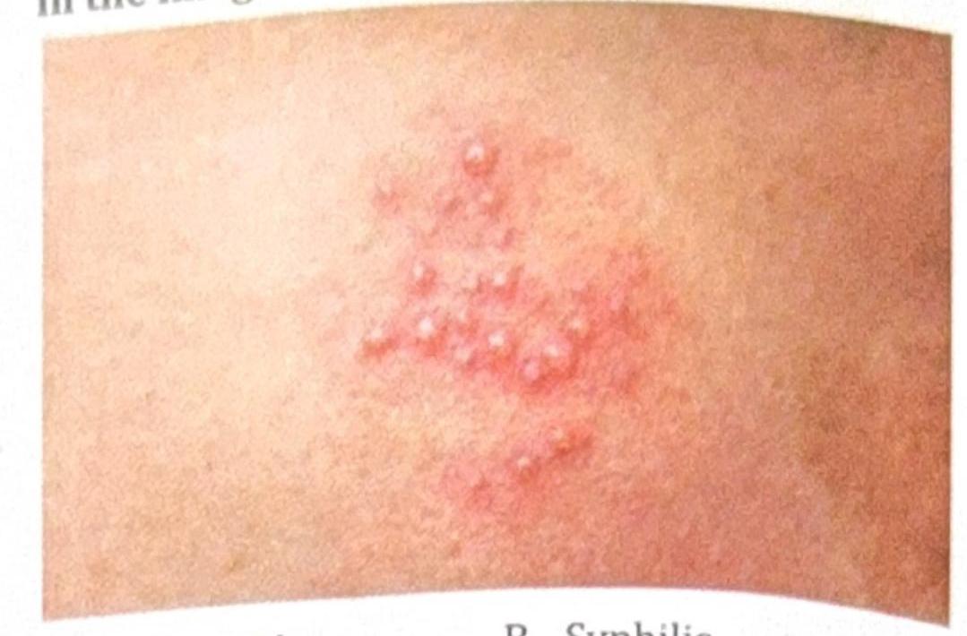
1 questionsA patient presents with painful vesicles as shown in the image. What is the diagnosis?
Internal Medicine
2 questionsA 40-year-old farmer presents with fever, calf tenderness, conjunctival suffusion, retro-orbital pain, and hypokalemia. What is the diagnosis?
A patient living with HIV presents with foulsmelling stools. Microscopic examination of the stool reveals no cysts or ova, but a 200-micrometer larva is observed. What is the most likely pathogen?
Microbiology
6 questionsA patient was admitted with bloody diarrhea after consumption of oysters. The organism exhibits the Kanagawa phenomenon. What is the correct organism?
Desert rheumatism is caused by:
Broad-based budding yeasts are seen in:
A female patient presents with dysuria and frequency. A coagulase-negative, novobiocin-resistant Staphylococcus species (>10^4 CFU/mL) was grown in urine culture. What does this indicate?
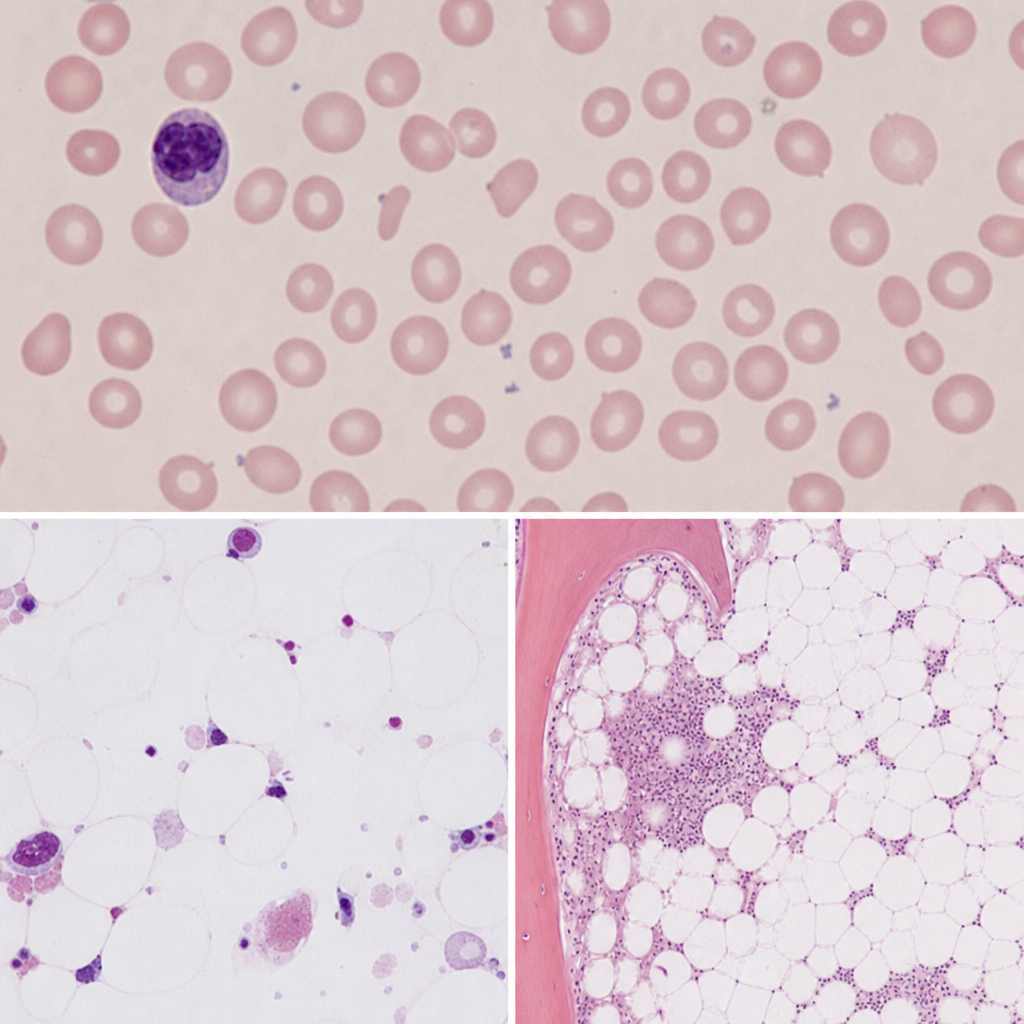
Which viral infection is most likely responsible for triggering this aplastic crisis in the patient?
A patient was diagnosed with Escherichia coli O157:H7 infection. In this designation, what does the "H" stand for?
Pediatrics
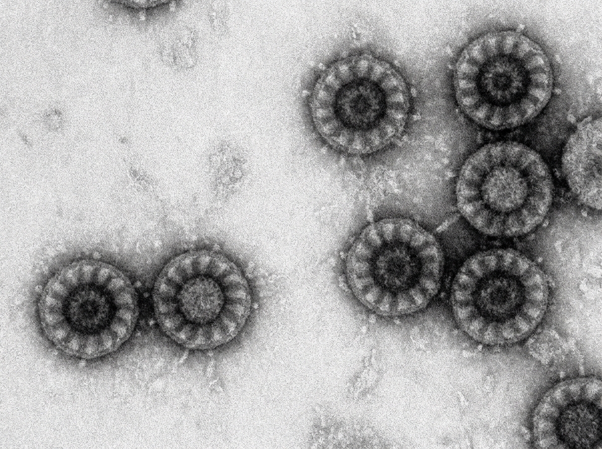
1 questionsA 2 year old child came with watery diarrhea. Electron Microscopy (EM) Image is shown here. Choose the correct pathogen.