Biochemistry
1 questionsWhich of the following statements is true regarding the functions of cAMP and cGMP?
Forensic Medicine
1 questionsWhich of the following is not a feature of postmortem staining?
Internal Medicine
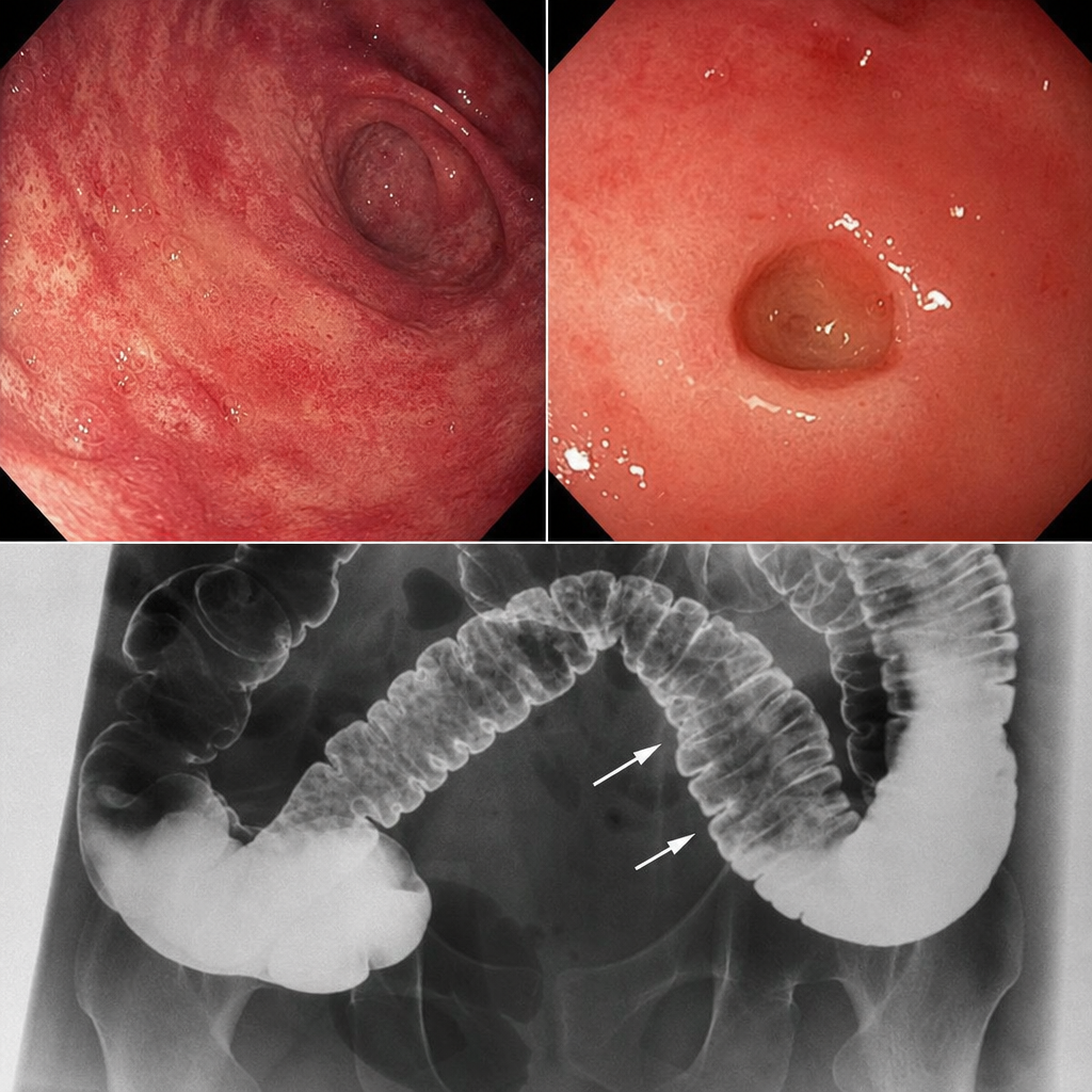
1 questionsA patient presents with skin involvement and collar stud ulceration in the intestine observed on radiography. What is the most likely diagnosis?
Pathology
3 questionsIgA nephropathy is not associated with which of the following?
Which of the following is a chromosomal instability syndrome?
Obliterative endarteritis in vasa vasorum is seen in -
Pharmacology
1 questionsWhich of the following is an action of muscarinic cholinergic receptors?
Physiology
3 questionsRebound increase in gastric acid secretion after stopping proton pump inhibitor therapy is due to?
'Flare' in Triple response is mediated by :
What is one of the specific functions of the primary motor cortex located on the anterior edge of the pre-central gyrus?