Forensic Medicine
1 questionsDuring a postmortem examination of a young adult found with a faded tattoo, relatives mentioned that the tattoo was once visible. What is the best method to identify the tattoo?
Internal Medicine
1 questionsA 50 year old male presents with fever and malaise for 4 months and pain in the knees and ankles. Blood tests are normal apart from a raised ESR. Chest x-ray shows bilateral hilar adenopathy and pulmonary infiltrates most severe in the upper and mid zones. Mantoux test is negative. What is the most likely diagnosis?
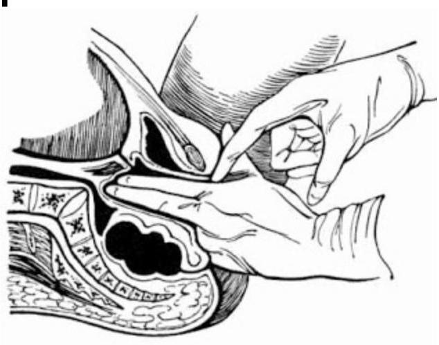
Obstetrics and Gynecology
1 questionsWhich of the following describes the points marked in the diagram of pelvic measurements?
Pharmacology
1 questionsWhich of the following actions is NOT associated with tricyclic antidepressants?
Psychiatry
5 questionsIn which condition is sex reassignment surgery typically performed?
Loosening of association is an example of
What is a common medical treatment for sexual paraphilias?
What is the primary cause of death in Neuroleptic Malignant Syndrome?
What type of disorder is Tourette syndrome?
Radiology
1 questionsInvestigation of choice for acute intracerebral hemorrhage is -