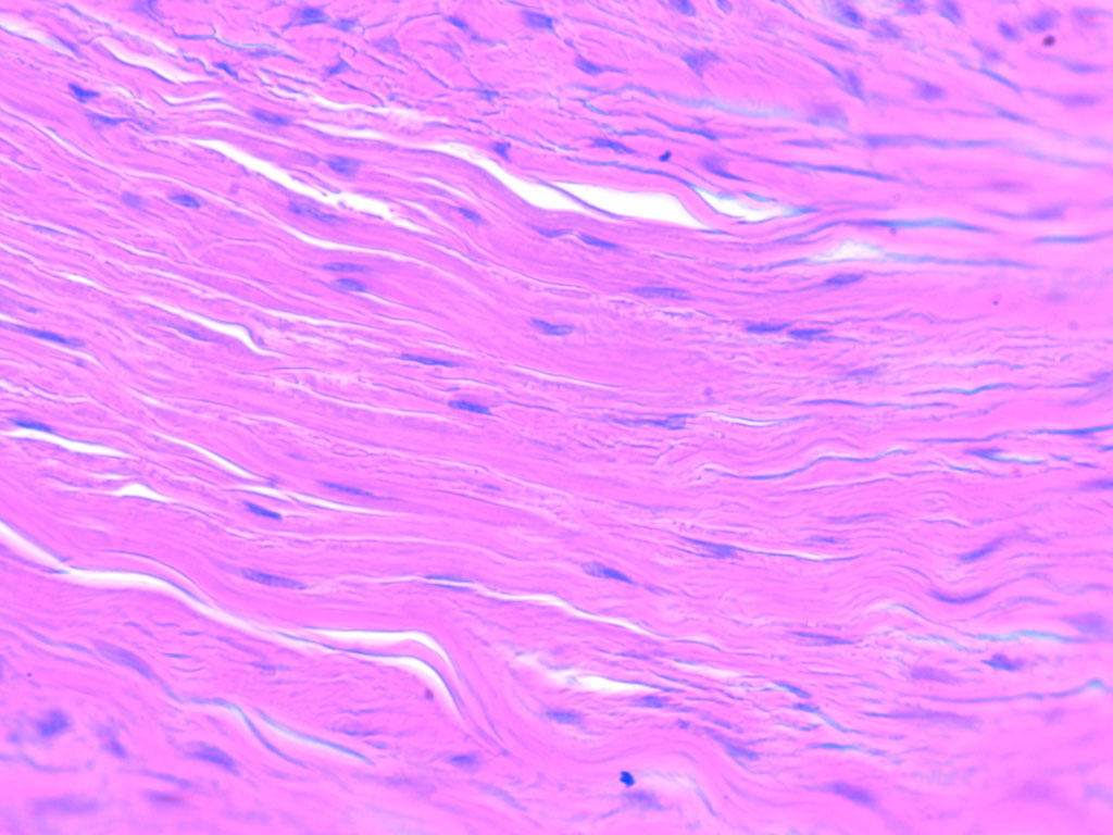
Anatomy
1 questionsUnable to consolidate long-term memory. Which lobe of the brain is involved?
NEET-PG 2012 - Anatomy NEET-PG Practice Questions and MCQs
Question 911: Unable to consolidate long-term memory. Which lobe of the brain is involved?
- A. Frontal
- B. Parietal
- C. Temporal (Correct Answer)
- D. Occipital
Explanation: ***Temporal*** - The **temporal lobe**, particularly the **hippocampus** within it, is critically involved in the formation and consolidation of new long-term memories [1]. - Damage to the temporal lobe can result in **anterograde amnesia**, the inability to form new memories after the injury [1]. *Frontal* - The **frontal lobe** is primarily responsible for executive functions, **planning**, decision-making, and working memory [1]. - While it contributes to memory retrieval and strategic memory processes, it is not the primary site for the consolidation of long-term memories. *Parietal* - The **parietal lobe** is involved in processing **sensory information**, spatial awareness, and navigation. - It plays a role in attention and short-term memory, but not in the consolidation of long-term memory. *Occipital* - The **occipital lobe** is dedicated to **visual processing** and interpretation of visual information. - It has no direct primary role in the consolidation of long-term memories.
Internal Medicine
7 questionsHypophosphatemia is seen in:
A pregnant woman is diagnosed with Graves' disease. The most appropriate therapy for her would be:
Chemotherapeutic Agent of Choice for the treatment of CML?
Best provocative test for diagnosis of Gastrinoma is:
What is the most common presentation for IgA nephropathy?
Fibrosis of upper lobe is due to
Which of the following is NOT a common pathogen causing pneumonia in COPD patients?
NEET-PG 2012 - Internal Medicine NEET-PG Practice Questions and MCQs
Question 911: Hypophosphatemia is seen in:
- A. Hyperthyroidism
- B. Hypoparathyroidism
- C. Hyperparathyroidism (Correct Answer)
- D. Pseudohypoparathyroidism
Explanation: ***Hyperparathyroidism*** - In **primary hyperparathyroidism**, the excess **parathyroid hormone (PTH)** leads to increased phosphate excretion by the kidneys [1], [4]. - This results in **hypophosphatemia** as the body attempts to maintain **calcium-phosphate balance**, often at the expense of phosphate levels [1]. *Hyperthyroidism* - While hyperthyroidism can affect **bone metabolism**, it is typically associated with **normal or slightly elevated phosphate levels**, not hypophosphatemia [3]. - The main electrolyte disturbances are usually related to **calcium** (e.g., hypercalcemia) due to increased bone turnover [3]. *Hypoparathyroidism* - **Hypoparathyroidism** is characterized by **low or absent PTH**, leading to decreased renal phosphate excretion. - This results in **hyperphosphatemia**, along with **hypocalcemia** [2]. *Pseudohypoparathyroidism* - In **pseudohypoparathyroidism**, there is **PTH resistance** at target tissues, even with high or normal PTH levels [2]. - This leads to symptoms resembling hypoparathyroidism, including **hyperphosphatemia** and **hypocalcemia** [2].
Question 912: A pregnant woman is diagnosed with Graves' disease. The most appropriate therapy for her would be:
- A. Radioiodine therapy
- B. Total thyroidectomy
- C. Carbimazole parenteral
- D. Propylthiouracil oral (Correct Answer)
Explanation: ***Propylthiouracil oral*** - **Propylthiouracil (PTU)** is the preferred antithyroid drug during the **first trimester** of pregnancy due to a lower risk of teratogenicity compared to methimazole/carbimazole [1]. - It works by inhibiting both the synthesis of thyroid hormones and the peripheral conversion of **T4 to T3**. *Radioiodine therapy* - **Radioactive iodine** is absolutely contraindicated in pregnancy as it can cross the placenta and cause **fetal hypothyroidism or athyreosis**. - It leads to permanent destruction of the thyroid gland and is not suitable for a temporary condition in a pregnant woman. *Total thyroidectomy* - While thyroidectomy can be considered for Graves' disease in pregnancy, it is generally reserved for cases where antithyroid drugs are not tolerated or ineffective, or for very large goiters causing compressive symptoms. - It carries risks associated with **surgery and anesthesia** during pregnancy, and requires **lifelong thyroid hormone replacement**. *Carbimazole parenteral* - **Carbimazole** (which is metabolized to methimazole) is generally avoided in the **first trimester** due to an increased risk of teratogenicity, particularly **aplasia cutis**, omphalocele, and choanal atresia [1]. - While it can be used in the second and third trimesters, **PTU is preferred in the first trimester**, and carbimazole is not typically administered parenterally.
Question 913: Chemotherapeutic Agent of Choice for the treatment of CML?
- A. Imatinib (Correct Answer)
- B. Vincristine
- C. Cyclophosphamide
- D. Methotrexate
Explanation: ***Imatinib*** - **Imatinib** is a **tyrosine kinase inhibitor** specifically targeting the **BCR-ABL fusion protein**, which is the hallmark of CML [1][2]. - It dramatically improved the prognosis of CML patients, making it the **first-line therapy** and agent of choice due to its high efficacy and relatively low toxicity compared to conventional chemotherapy [2]. *Vincristine* - **Vincristine** is a **vinca alkaloid** that inhibits microtubule formation, primarily used in acute leukemias and lymphomas. - It is not the agent of choice for CML due to its different mechanism of action and the availability of more targeted therapies for CML. *Cyclophosphamide* - **Cyclophosphamide** is an **alkylating agent** that causes DNA damage, used in various cancers and autoimmune diseases. - While it can be used in some hematologic malignancies, it is not the preferred or most effective treatment for CML, especially given the success of targeted therapies. *Methotrexate* - **Methotrexate** is an **antimetabolite** that interferes with DNA synthesis, commonly used in acute leukemias, lymphomas, and autoimmune conditions. - It is not considered the chemotherapeutic agent of choice for CML, as its mechanism of action is not specific to the BCR-ABL anomaly characteristic of CML.
Question 914: Best provocative test for diagnosis of Gastrinoma is:
- A. Ca++ infusion test
- B. Secretin injection test (Correct Answer)
- C. ACTH stimulation test
- D. Steroid assay
Explanation: ***Secretin injection test*** - The **secretin injection test** is the most reliable provocative test for gastrinoma, leading to a paradoxical increase in gastrin levels [1]. - In normal individuals, secretin suppresses gastrin release, but in gastrinoma, it stimulates **gastrin secretion** [1]. *Ca++ infusion test* - The **calcium infusion test** can also stimulate gastrin release in gastrinoma patients, but it is less specific and potentially more risky than the secretin test due to potential side effects like cardiac arrhythmias. - It involves infusing calcium gluconate to observe any uncharacteristic rise in gastrin. *ACTH stimulation test* - The **ACTH stimulation test** is used to evaluate adrenal gland function, particularly in suspected cases of adrenal insufficiency or hypercortisolism [2]. - It does not have any direct relevance to the diagnosis of **gastrinoma**. *Steroid assay* - **Steroid assays** measure levels of various steroid hormones (e.g., cortisol, aldosterone) in the body to assess adrenal or gonadal function. - This test is not used for diagnosing **gastrinoma**.
Question 915: What is the most common presentation for IgA nephropathy?
- A. Nephritic syndrome
- B. Nephrotic syndrome
- C. Microscopic hematuria
- D. Repeated gross hematuria (Correct Answer)
Explanation: ***Repeated gross hematuria*** - The hallmark of **IgA nephropathy** is recurrent episodes of **gross hematuria**, particularly following **respiratory infections** [1]. - It is often associated with **renal impairment** but can present initially with **visible blood** in the urine [1]. *Nephritic syndrome* - While IgA nephropathy can lead to nephritic features, it does not commonly present primarily as **nephritic syndrome**, which includes hypertension and edema. - Nephritic syndrome is characterized by significant **proteinuria** and acute renal failure, rather than the classic presentation of hematuria [2]. *Microscopic hematuria* - Although **microscopic hematuria** can occur in IgA nephropathy, it is not the most common and noticeable presentation; **gross hematuria** is more characteristic [1]. - Microscopic hematuria lacks the acute visual symptoms seen in cases proving the diagnosis. *Nephritic syndrome* - This option is a repetition of and does not provide any additional unique characteristics specific to **IgA nephropathy**. - It shares the same clinical features discussed previously and is thus not representative of the most common presentation.
Question 916: Fibrosis of upper lobe is due to
- A. ABPA (Correct Answer)
- B. Rheumatoid arthritis
- C. Certain types of pneumonia
- D. Bronchiectasis
Explanation: ***ABPA*** - **Allergic bronchopulmonary aspergillosis (ABPA)** is a hypersensitivity reaction to *Aspergillus* species, particularly *A. fumigatus*, which colonize the airways, and commonly leads to **upper lobe fibrosis** and **bronchiectasis**. - The chronic inflammation and recurrent immune responses result in progressive airway damage, characterized on imaging by centrilobular nodules, mucoid impaction, and ultimately **fibrosis in the upper lobes**. Other conditions causing similar upper lobe conglomerate masses include Progressive Massive Fibrosis (PMF) seen in coal worker's pneumoconiosis [1]. *Certain types of pneumonia* - While pneumonia can cause inflammation, it typically leads to **lobar consolidation** or diffuse infiltrates rather than specific upper lobe fibrosis. - **Resolution of pneumonia** usually occurs without significant fibrotic changes, unlike chronic conditions that predispose to fibrosis. *Bronchiectasis* - **Bronchiectasis** is a general term for permanent dilation of the bronchi, which can occur in any lobe, but alone **does not directly cause upper lobe fibrosis** as a primary etiology. - While it often coexists with conditions that cause fibrosis (like ABPA or CF), it's a consequence of airway damage, not the direct cause of the fibrotic process itself; it can however predispose to recurrent infections leading to scarring. *Rheumatoid arthritis* - **Rheumatoid arthritis** can cause interstitial lung disease, including **pulmonary fibrosis**, but it typically manifests as a **basilar or diffuse pattern** rather than predominantly upper lobe fibrosis [2]. - The fibrosis associated with RA-ILD is commonly of a **usual interstitial pneumonia (UIP)** or **non-specific interstitial pneumonia (NSIP)** pattern [2].
Question 917: Which of the following is NOT a common pathogen causing pneumonia in COPD patients?
- A. Klebsiella pneumoniae
- B. Legionella spp (Correct Answer)
- C. Pseudomonas aeruginosa
- D. Haemophilus influenzae
Explanation: ***Legionella spp*** - While *Legionella* can cause pneumonia, it is **not a common pathogen** specifically in COPD exacerbations or community-acquired pneumonia in these patients [1]. - *Legionella* pneumonia often presents with **extrapulmonary symptoms** like diarrhea and hyponatremia and is typically associated with contaminated water sources [1]. *Haemophilus influenzae* - This is a very common pathogen causing both **acute exacerbations of COPD (AECOPD)** and pneumonia in patients with underlying COPD [1]. - COPD patients often have **impaired mucociliary clearance** and altered airway microbiology, making them susceptible to *H. influenzae* colonization and infection [1]. *Pseudomonas aeruginosa* - *Pseudomonas aeruginosa* is an important pathogen in **severe COPD exacerbations**, especially in patients with frequent exacerbations, bronchiectasis, or prior antibiotic use. - Its presence often indicates a **more severe disease course** and requires specific antibiotic coverage. *Klebsiella pneumoniae* - *Klebsiella pneumoniae* is a significant cause of **pneumonia in immunocompromised individuals**, including those with COPD, diabetes, or alcoholism. - It often leads to **severe, necrotizing pneumonia**, particularly in the upper lobes, and can cause abscess formation.
Pharmacology
1 questionsWhat is the initial drug of choice for a suspected case of acute adrenal insufficiency?
NEET-PG 2012 - Pharmacology NEET-PG Practice Questions and MCQs
Question 911: What is the initial drug of choice for a suspected case of acute adrenal insufficiency?
- A. Norepinephrine
- B. Hydrocortisone (Correct Answer)
- C. Dexamethasone
- D. Fludrocortisone
Explanation: ***Hydrocortisone*** - This is the initial drug of choice due to its **combined mineralocorticoid and glucocorticoid activity**, which effectively replaces the deficient hormones in acute adrenal insufficiency. - It also has a **rapid onset of action** crucial for stabilizing patients in an adrenal crisis. *Norepinephrine* - This is a **vasopressor** used to manage **severe hypotension or shock** by increasing peripheral vascular resistance. - While hypotension is a feature of adrenal insufficiency, norepinephrine does not address the underlying hormonal deficiency directly and is not the primary treatment. *Dexamethasone* - Dexamethasone is a **potent glucocorticoid** and can be used in adrenal insufficiency, but it lacks significant **mineralocorticoid activity**. - Its longer half-life might make it less ideal for immediate, titratable replacement compared to hydrocortisone in an acute setting. *Fludrocortisone* - Fludrocortisone is a **pure mineralocorticoid** primarily used for long-term replacement therapy to manage sodium and potassium balance in adrenal insufficiency. - It does not have sufficient glucocorticoid activity to address the immediate, life-threatening aspects of acute adrenal crisis.
Physiology
1 questionsLesion of globus pallidus causes
NEET-PG 2012 - Physiology NEET-PG Practice Questions and MCQs
Question 911: Lesion of globus pallidus causes
- A. Chorea
- B. Athetosis (Correct Answer)
- C. Hemibalismus
- D. Dystonia
Explanation: ***Athetosis*** - **Athetosis** is the **classic movement disorder** associated with lesions of the **globus pallidus**, often occurring with **putamen** involvement. - It is characterized by **slow, writhing, involuntary movements**, particularly affecting the **distal extremities** (hands and feet). - Commonly seen in **kernicterus** (bilirubin-induced damage to basal ganglia), **cerebral palsy**, and **status marmoratus** of the basal ganglia. - When combined with chorea, it forms **choreoathetosis**. *Chorea* - **Chorea** is predominantly associated with dysfunction of the **caudate nucleus** and **putamen**, as seen in **Huntington's disease**. - It involves brief, irregular, unpredictable, **involuntary movements** that flow from one body part to another. *Hemibalismus* - **Hemibalismus** is most commonly caused by a lesion in the **subthalamic nucleus** (nucleus of Luys), often due to a **lacunar stroke**. - It involves large-amplitude, **involuntary flinging movements** of the limbs on **one side of the body**. *Dystonia* - **Dystonia** involves sustained or repetitive muscle contractions leading to twisting movements or abnormal fixed postures. - It results from dysfunction of **multiple basal ganglia structures** including the globus pallidus internal segment, putamen, and thalamus, but is **not the classic presentation** of isolated globus pallidus lesions.