All (1216)Anatomy (104)Anesthesiology (21)Biochemistry (179)Community Medicine (104)Dental (9)Dermatology (21)ENT (2)Forensic Medicine (41)General Medicine (2)Internal Medicine (79)Microbiology (83)Obstetrics and Gynecology (63)Ophthalmology (68)Orthopaedics (36)Pathology (82)Pediatrics (43)Pharmacology (85)Physiology (91)Psychiatry (2)Psychiatry (20)Radiology (28)Surgery (53)
Anatomy
6 questionsQ191
What is the lower limit of the retropharyngeal space?
Q192
Which is the primary segment of the liver drained by the right hepatic vein?
Q193
In walking, gravity tends to tilt pelvis and trunk to the unsupported side, the major factor in preventing this unwanted movement is?
Q194
Where is the neurovascular plane located in the anterior abdominal wall?
Q195
The thyrocervical trunk is a branch of which part of subclavian artery?
Q196
Which of the following cell types is neuroectodermal in origin?
Biochemistry
2 questionsQ191
Which macronutrient has the highest thermogenic effect?
Q192
What coenzyme is required by gulonate dehydrogenase for its activity?
Pathology
1 questionsQ191
Which condition is most commonly associated with systemic amyloidosis?
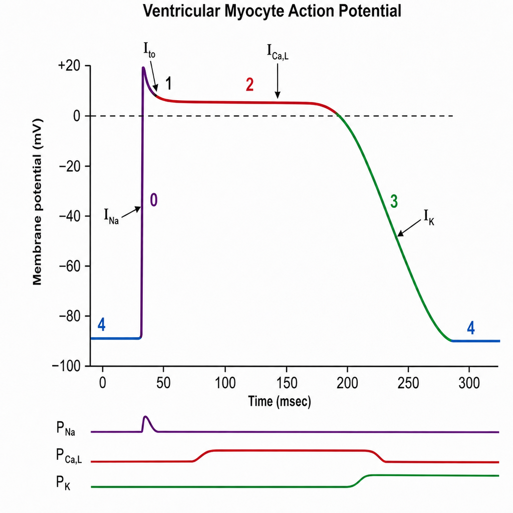
Physiology
1 questionsQ191
The plateau phase of this graph is due to: