All (113)Anatomy (3)Community Medicine (31)ENT (1)Forensic Medicine (1)Internal Medicine (5)Microbiology (1)Obstetrics and Gynecology (30)Ophthalmology (1)Orthopaedics (2)Pathology (3)Pediatrics (4)Physiology (1)Psychiatry (1)Radiology (1)Surgery (28)
Q21
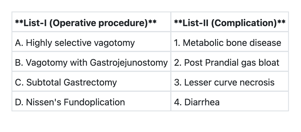
Match List-I with List-II and select the correct answer using the code given below the Lists: **List-I (Procedure)** A. Highly selective vagotomy B. Vagotomy with gastrojejunostomy C. Subtotal gastrectomy D. Nissen's fundoplication **List-II (Complication)** 1. Metabolic bone disease 2. Post-prandial gas bloat 3. Lesser curve necrosis 4. Diarrhea **Code:**
Q22
Optimum age for surgery for a child with cleft lip is:
Q23
An electrical contact burn is considered to be:
Q24
A 52 year old male patient comes with history of rectal bleeding, alteration in bowel habits and tenesmus. The ideal investigation would be:
Q25
What is the treatment of choice in a patient with Crohn’s disease, where inflamed appendix was found on exploration?
Q26
The following are features of hypovolemic shock except:
Q27
A 45 year old man sustains trauma in a road traffic accident and develops engorgement of neck veins, pallor, rapid pulse rate, and chest pain. What is the most likely diagnosis?