INI-CET 2018 — Radiology
5 Previous Year Questions with Answers & Explanations
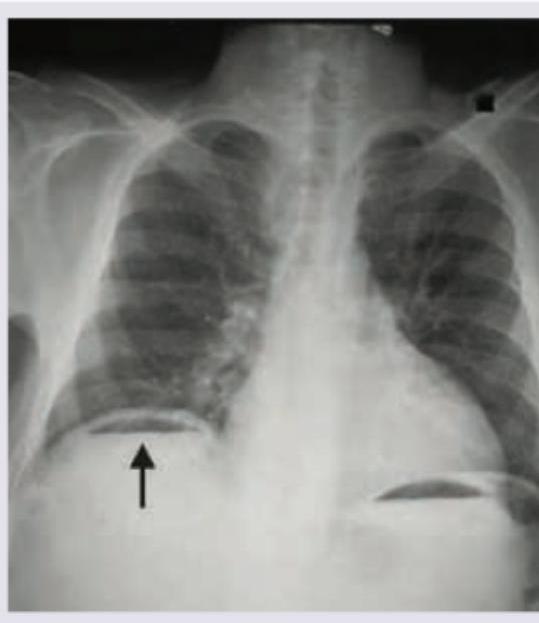
What does the given chest X-ray show?
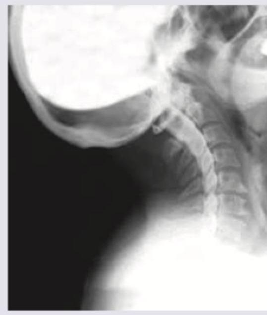
Identify the type of investigation shown in the image below.
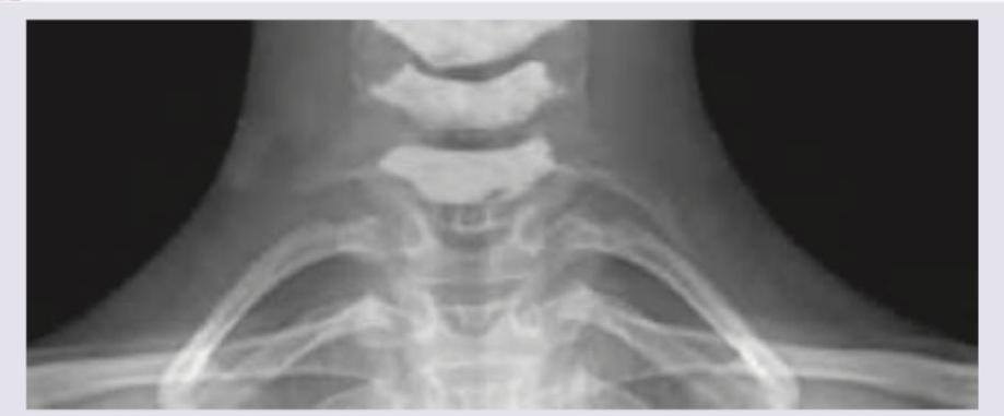
A 25-year-old female presents with neck pain and tingling sensation in her left arm. An X-ray of the cervicothoracic region is obtained. What is the radiological finding shown in the image?
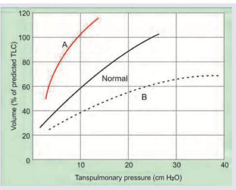
Curve A signifies which of the following?
Identify the lesion in a 20-year-old male whose foot X-ray is shown below:
INI-CET 2018 - Radiology INI-CET Practice Questions and MCQs
Question 1: What does the given chest X-ray show?
- A. Pneumoperitoneum (Correct Answer)
- B. Emphysema
- C. Diaphragmatic hernia
- D. Diaphragmatic eventration
Explanation: ***Pneumoperitoneum*** - The chest X-ray shows **free air under the diaphragm**, visible as a lucent (dark) crescent between the liver/spleen and the diaphragm (indicated by the arrow on the right side of the patient). - This finding is diagnostic of **pneumoperitoneum**, which is often caused by a perforated abdominal viscus like a peptic ulcer or bowel perforation. *Emphysema* - **Emphysema** is a lung condition characterized by over-inflated alveoli and air trapping within the lungs, leading to hyperlucency of the lung fields and flattened diaphragms. - It does not present as free air below the diaphragm but rather as changes within the lung parenchyma. *Diaphragmatic hernia* - A **diaphragmatic hernia** involves the protrusion of abdominal organs into the chest cavity through a defect in the diaphragm. - This would typically show abdominal contents (e.g., bowel loops or stomach) above the diaphragm in the thoracic cavity, not free air below it. *Diaphragmatic eventration* - **Diaphragmatic eventration** is an abnormal elevation of part or all of an intact hemidiaphragm due to thinning and weakness of the diaphragmatic muscle. - It causes an elevated diaphragm but does not involve free air in the peritoneal cavity.
Question 2: Identify the type of investigation shown in the image below.
- A. Angiography
- B. Myelography (Correct Answer)
- C. Neurography
- D. Fluoroscopy
Explanation: ***Correct: Myelography*** - The image displays **contrast agent within the spinal canal**, outlining the spinal cord and nerve roots against the bony structures of the cervical spine - This technique is used to visualize **nerve impingement, disc herniation, or spinal cord compression** - Characteristic finding: contrast delineating the thecal sac and nerve root sleeves *Incorrect: Angiography* - Angiography involves injecting contrast into **blood vessels** to visualize vascular structures, detect blockages, or aneurysms - The image shows the **spinal canal** rather than the vascular tree *Incorrect: Neurography* - Neurography (MR neurography) is a specialized **MRI technique** to visualize peripheral nerves themselves - Does not involve injection of contrast into the spinal canal as shown in the image *Incorrect: Fluoroscopy* - Fluoroscopy is a **real-time X-ray imaging technique** used for dynamic assessment or procedure guidance - While fluoroscopy may be used **during** myelography to guide needle placement, the specific technique of contrast visualization in the spinal canal defines this as myelography
Question 3: A 25-year-old female presents with neck pain and tingling sensation in her left arm. An X-ray of the cervicothoracic region is obtained. What is the radiological finding shown in the image?
- A. Cervical rib (Correct Answer)
- B. Costochondritis
- C. Fracture of 2nd rib
- D. Spondylolisthesis
Explanation: ***Cervical rib*** - The image displays an extra rib arising from the **C7 cervical vertebra**, which is characteristic of a cervical rib. - This **supernumerary rib** extends towards the sternum or first thoracic rib, a classic radiological finding. *Costochondritis* - **Costochondritis** is an inflammation of the cartilage connecting the ribs to the sternum, which is typically a clinical diagnosis, not visible on X-ray. - An X-ray would not show inflammatory changes in cartilage or soft tissue, making this diagnosis unlikely based on imaging alone. *Fracture of 2nd rib* - A **fracture of the 2nd rib** would appear as a discontinuity or break in the normal bony architecture of the second rib. - The image does not show any signs of a broken rib; instead, it shows an **extra, well-formed rib-like structure** originating from the cervical spine. *Spondylolisthesis* - **Spondylolisthesis** involves the anterior displacement of one vertebral body over another, usually in the lumbar spine. - This condition is also not visible in the provided image, which focuses on the cervicothoracic junction and shows an **anatomic variation** rather than vertebral slippage.
Question 4: Curve A signifies which of the following?
- A. (a) (Correct Answer)
- B. (b)
- C. (c)
- D. (d)
Explanation: ***Option (a) - Emphysema (Curve A is correct)*** - Curve A shows a **leftward shift** compared to the normal curve on the pressure-volume diagram - For any given transpulmonary pressure, a **higher lung volume** is achieved - This indicates **increased lung compliance** - the lungs are easier to inflate - Characteristic of **emphysema**, where there is **loss of elastic recoil** due to destruction of alveolar walls and elastic tissue - In emphysema, lungs inflate easily but have difficulty deflating due to loss of elastic recoil *Option (b) - Normal* - This would represent the **baseline normal pressure-volume curve** - Serves as a reference point to compare pathological states - Shows normal lung compliance and elastic recoil *Option (c) - Pulmonary Fibrosis* - This would show a **rightward shift** on the pressure-volume curve - For any given transpulmonary pressure, a **lower lung volume** is achieved - Indicates **decreased lung compliance** - the lungs are stiffer and harder to inflate - Characteristic of **restrictive lung diseases** like pulmonary fibrosis, where excessive collagen deposition makes lungs stiff *Option (d) - Other Pathological State* - This would represent another abnormal curve pattern - Could include conditions like ARDS, pneumothorax, or other restrictive/obstructive patterns - The specific interpretation depends on the curve shown in the image
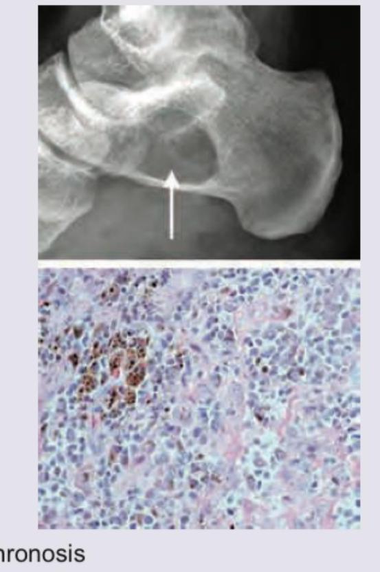
Question 5: Identify the lesion in a 20-year-old male whose foot X-ray is shown below:
- A. Ochronosis
- B. Haemophilic pseudo-tumour
- C. Pigmented villonodular synovitis
- D. Mycetoma (Correct Answer)
Explanation: ***Mycetoma*** - The X-ray image shows a **lytic lesion** with surrounding sclerosis in the calcaneus, indicated by the arrow, which is characteristic of **mycetoma**. The histological image reveals an inflammatory infiltrate with **pigmented fungal grains (brown aggregates)**, confirming the diagnosis. - Mycetoma is a chronic granulomatous infection of subcutaneous tissues, often extending to bone, particularly in the foot. Its characteristic features include **grains (colonies of microorganisms)** within the lesions. *Ochronosis* - Ochronosis would show **dark discoloration of cartilage** and connective tissues due to **homogentisic acid** deposition, leading to degenerative arthritis. This would appear on X-ray as **calcification of cartilage** (e.g., intervertebral discs, menisci) rather than focal lytic lesions with grains. - The histological image would show characteristic **ochre-colored pigment deposits**, not fungal grains. *Haemophilic pseudo-tumour* - A haemophilic pseudo-tumour is a rare complication of hemophilia, often presenting as a **large, expanding lesion** within bone or soft tissue, typically due to recurrent hemorrhage. - X-rays would reveal a **well-defined, expansile lesion** with cortical thinning or bone destruction, and histology would show features of chronic hemorrhage, fibrous tissue, and hemosiderin deposition, not fungal grains. *Pigmented villonodular synovitis* - This condition involves **proliferation of synovial tissue**, often affecting large joints like the knee or hip, leading to bone erosions and cysts. - Histology would show **hyperplastic synovium** with hemosiderin deposition, multinucleated giant cells, and lipid-laden macrophages, but not fungal grains as seen in the microscopy image.