All (71)Anatomy (1)Anatomy (22)Biochemistry (1)Community Medicine (3)Dermatology (4)Forensic Medicine (8)Microbiology (4)Ophthalmology (3)Orthopaedics (4)Pathology (5)Pathology (2)Pediatrics (1)Physiology (2)Psychiatry (1)Radiology (3)Surgery (4)Surgery (3)
Q61
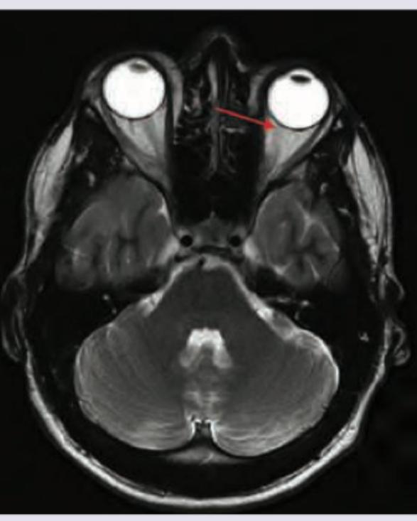
The marked extraocular muscle has a cranial nerve nucleus. At what level in the brain is the nucleus located? (AIIMS May 2018)
Q62
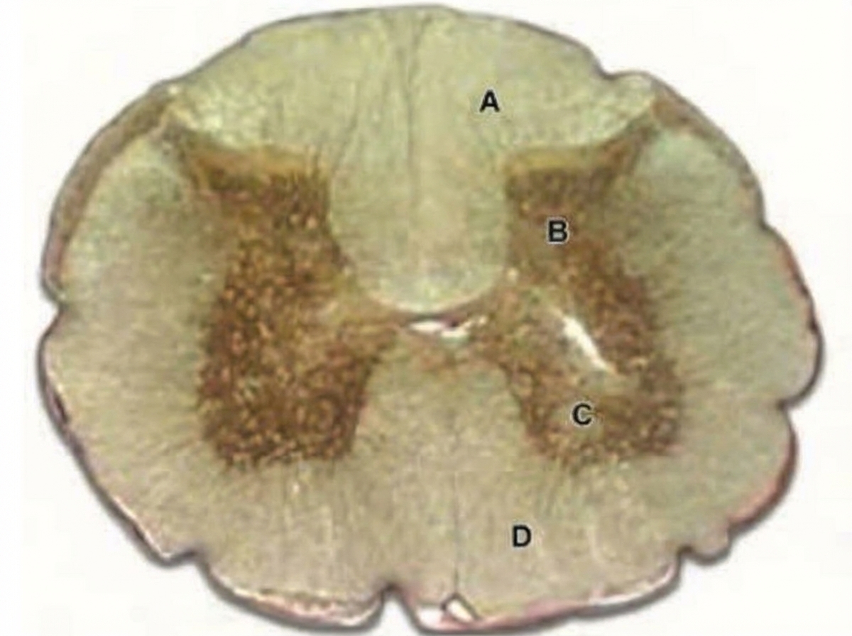
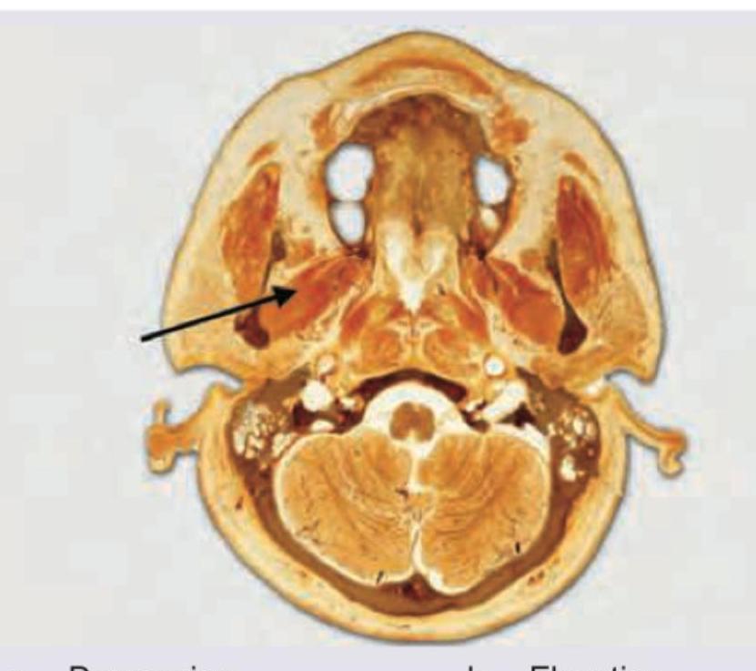
What is the function of the muscle marked in the cut section shown below? (AIIMS May 2018)
Q63
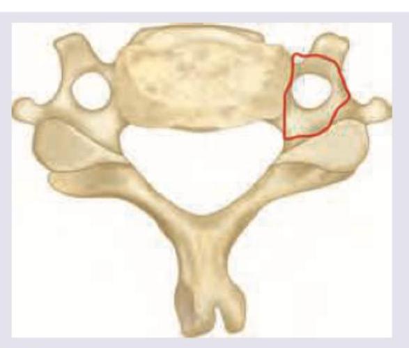
Which structure passes through the area marked in red? (AIIMS May 2018)
Q64
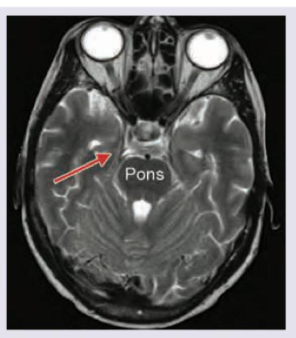
Which structure is marked with a red arrow in the image shown below? (AIIMS May 2018)
Q65
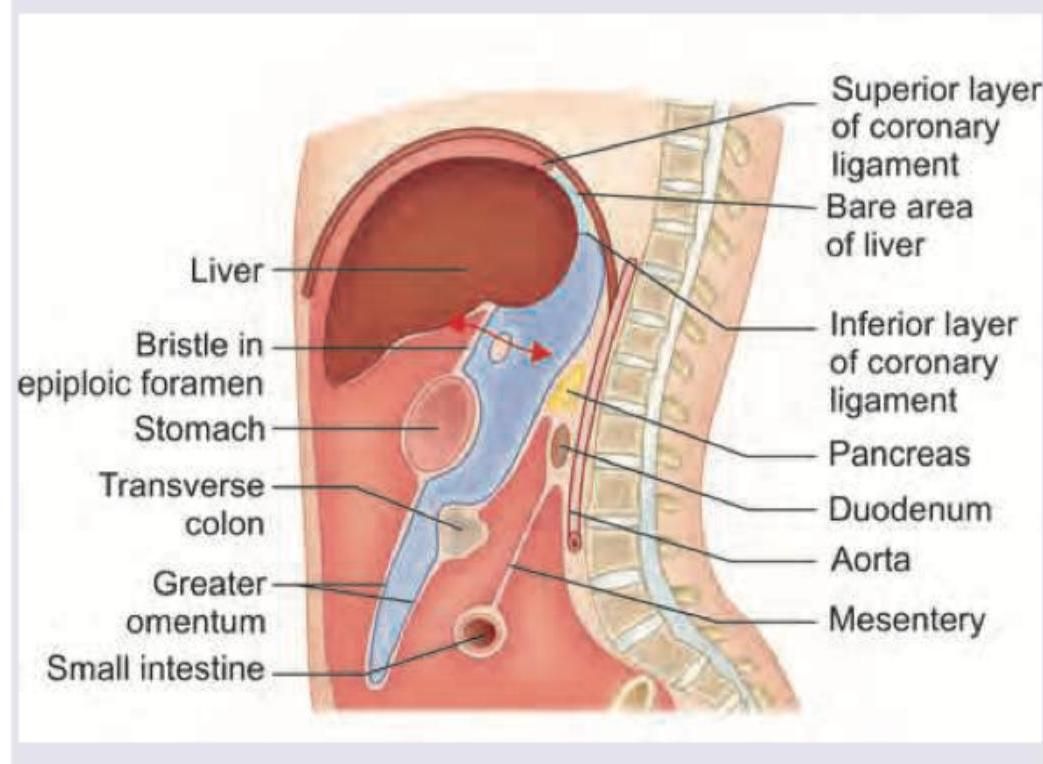
Identify the structure that forms the superior border of the epiploic foramen (marked in red) in the image below.
Q66
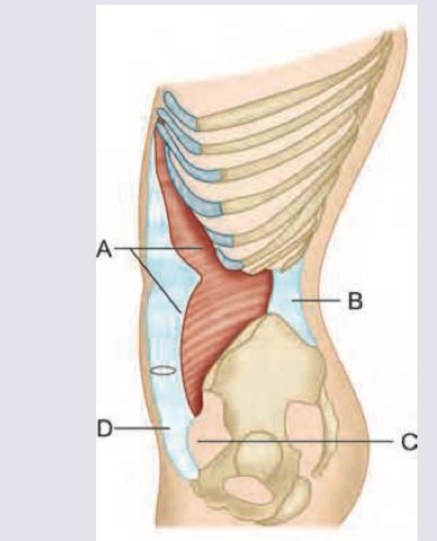
Which of the following marks the conjoint tendon?
Q67
Identify the urogenital diaphragm in the image given below:
Q68
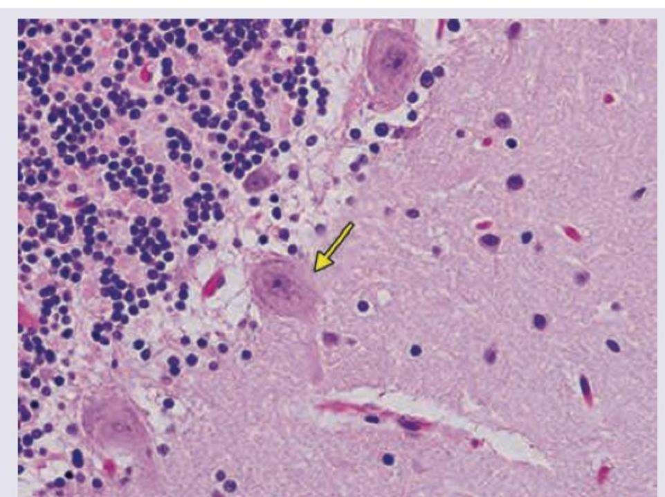
In the given slide of cerebellum, the marked cell is inhibitory to?