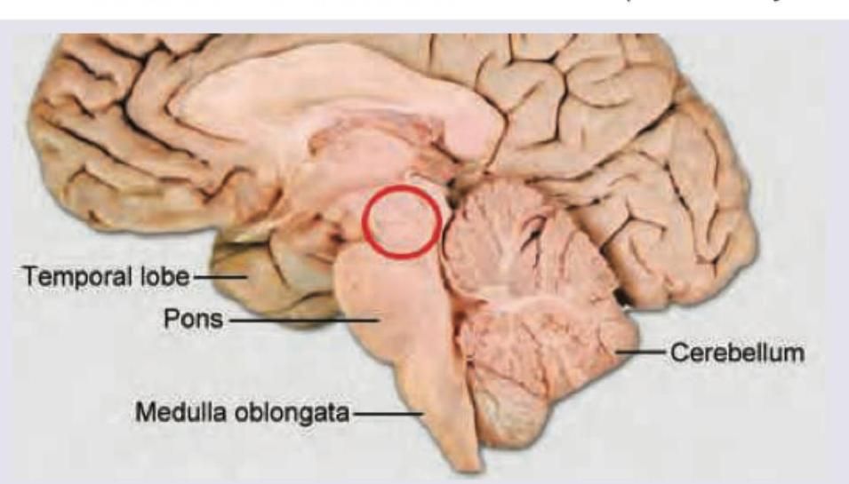
Q51
Identify the marked layer in the given histological section.
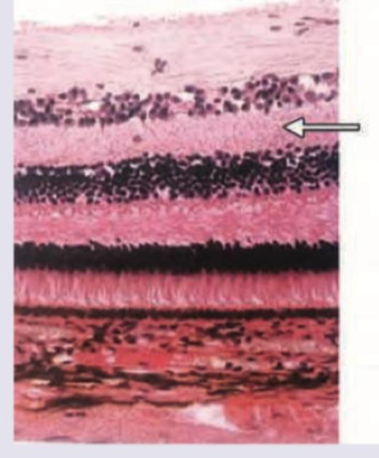
Q52
Identify the type of connective tissue present in the area marked with the arrow.
Q53
What is the action of the muscle marked in the given image?
Q54
Identify the part of the duodenum marked below:
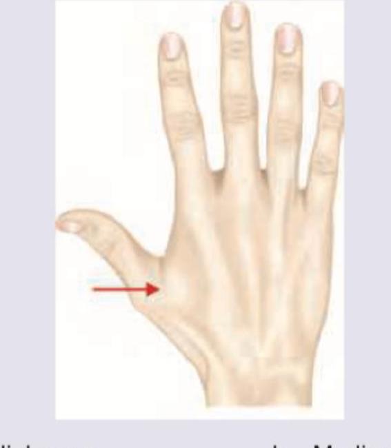
Q55
Which nerve gives sensory supply to the region marked with an arrow?
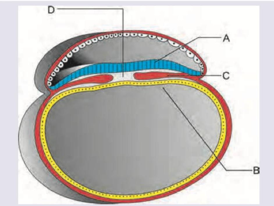
Q56
Nucleus pulposus develops from: (AIIMS May 2018)
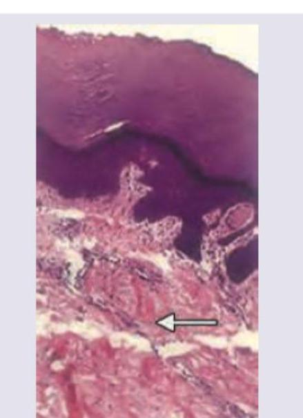
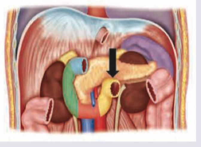
Q57
A young patient with absent thymus presents with tetany and hypoparathyroidism. Which of the following is a marked area in the picture defective in this condition?
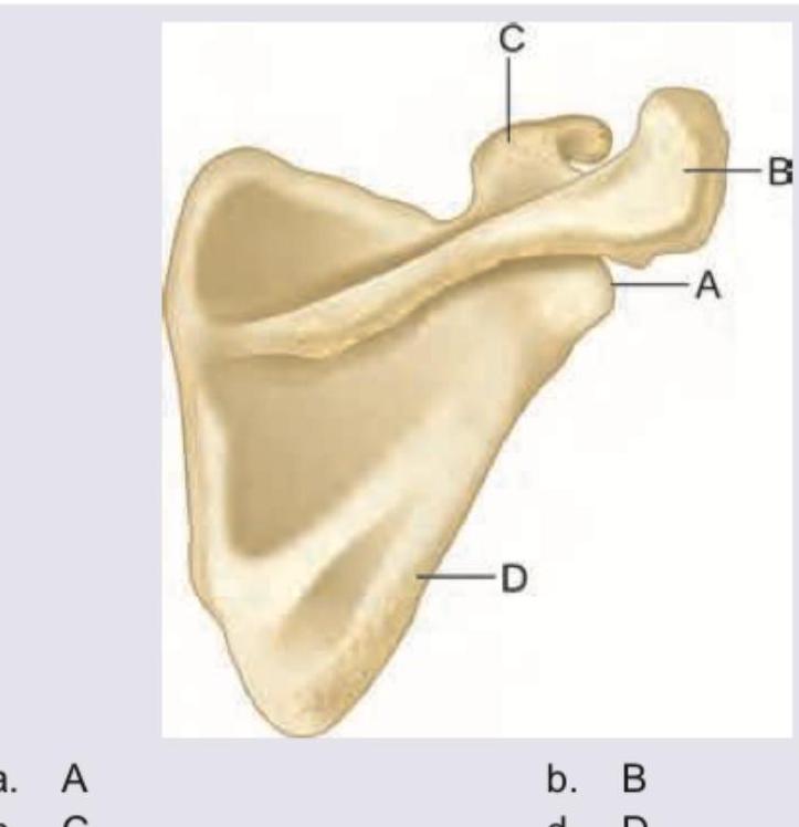
Q58
Which of the following structures of the scapula is palpable in infraclavicular fossa?
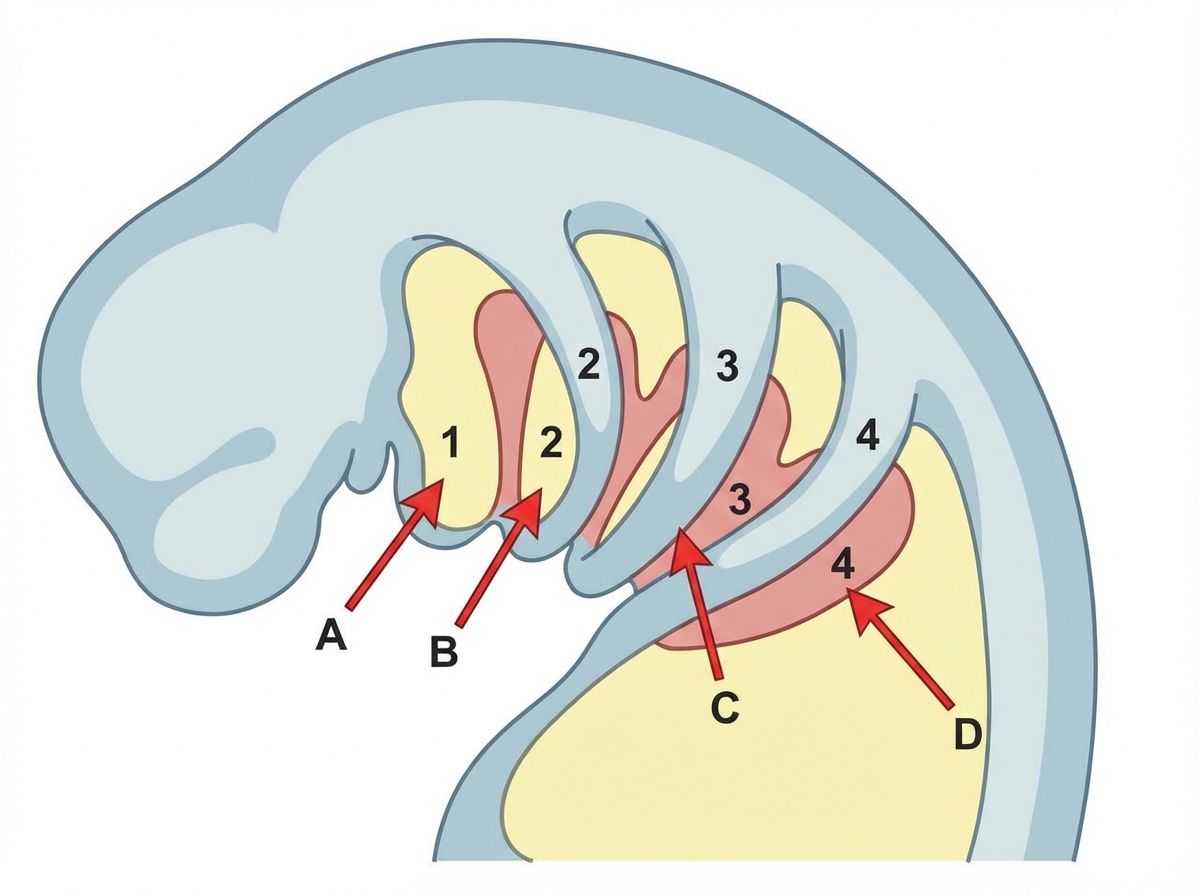
Q59
Mandibular division of trigeminal nerve passes through which of the following? (AIIMS May 2018)
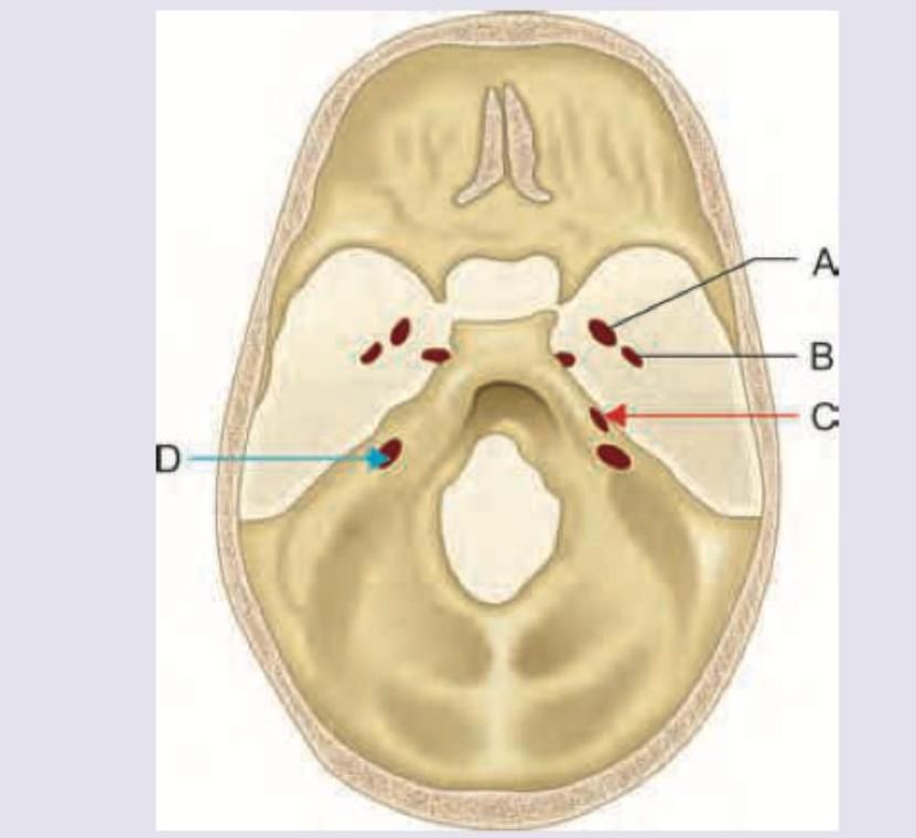
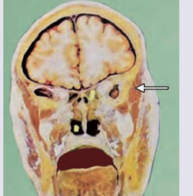
Q60
Which disease occurs due to involvement of the structure marked in red? (AIIMS May 2018)