All (71)Anatomy (1)Anatomy (22)Biochemistry (1)Community Medicine (3)Dermatology (4)Forensic Medicine (8)Microbiology (4)Ophthalmology (3)Orthopaedics (4)Pathology (5)Pathology (2)Pediatrics (1)Physiology (2)Psychiatry (1)Radiology (3)Surgery (4)Surgery (3)
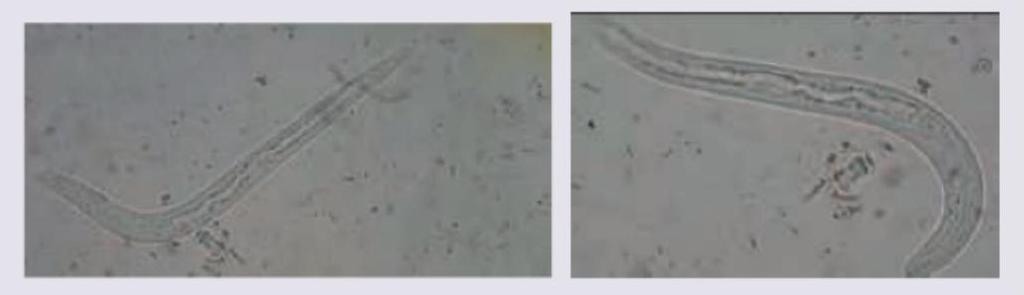
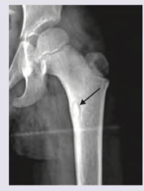
Q31
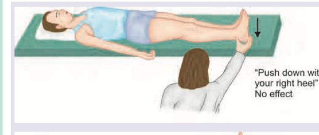
Which is incorrect about the image shown below?

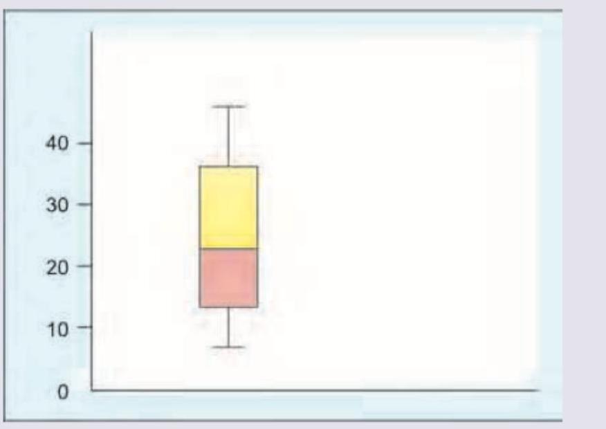
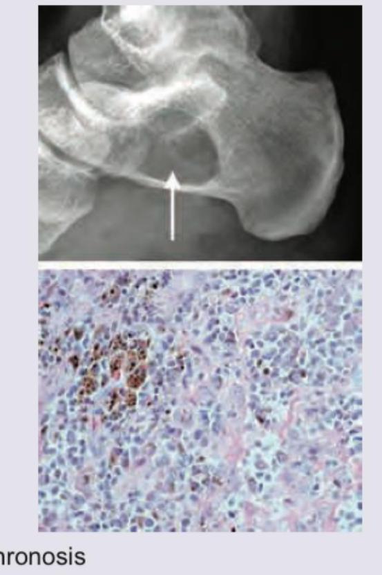
Q32
Which of the following is true about the box plot shown?