All (71)Anatomy (1)Anatomy (22)Biochemistry (1)Community Medicine (3)Dermatology (4)Forensic Medicine (8)Microbiology (4)Ophthalmology (3)Orthopaedics (4)Pathology (5)Pathology (2)Pediatrics (1)Physiology (2)Psychiatry (1)Radiology (3)Surgery (4)Surgery (3)
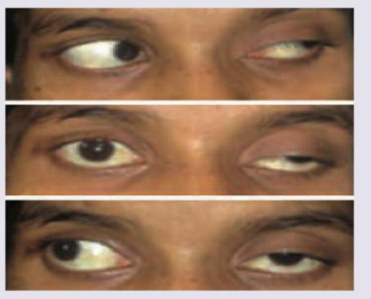
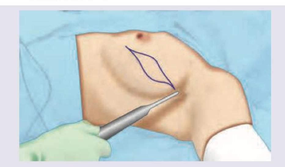
Q21
What is the best management of the case shown?
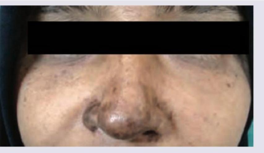
Q22
A patient presented with fever and joint pain for which she was put on NSAIDs. After 10 days she developed a skin lesion as shown in the image. Diagnosis is: