INI-CET 2018 — Forensic Medicine
7 Previous Year Questions with Answers & Explanations
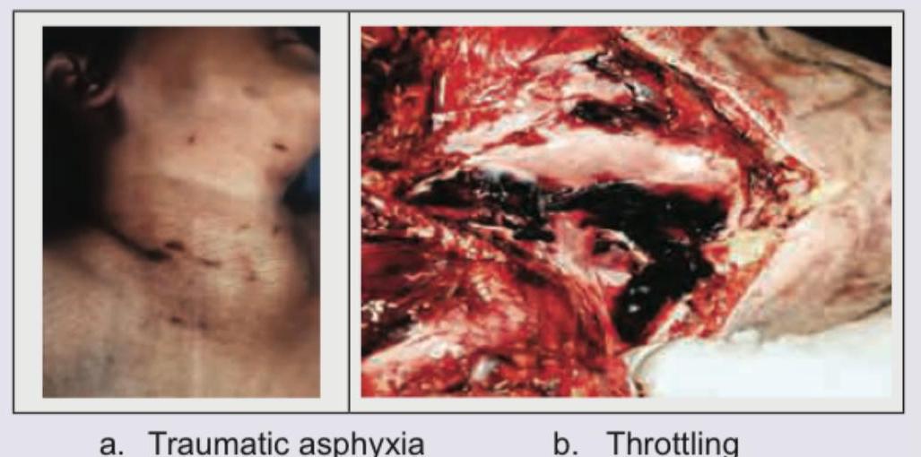
A female was found dead in her bedroom. The room was not locked from inside. Her blood alcohol value was found to be 350 mg/dL. The picture taken at the post mortem is shown below. The diagnosis is? (AIIMS Nov 2018, AIIMS Nov 2017)
The following image shows: (AIIMS Nov 2018)
The following presentation occurs due to? (AIIMS Nov 2018)
Identify the post mortem lividity of the image: (AIIMS Nov 2018)

Identify the flowers shown below:
Identify the correct pair: i. Mummification ii. Adipocere iii. Marbling iv. Livor mortis
Comment on age of the male patient:
INI-CET 2018 - Forensic Medicine INI-CET Practice Questions and MCQs
Question 1: A female was found dead in her bedroom. The room was not locked from inside. Her blood alcohol value was found to be 350 mg/dL. The picture taken at the post mortem is shown below. The diagnosis is? (AIIMS Nov 2018, AIIMS Nov 2017)
- A. Traumatic asphyxia
- B. Throttling (Correct Answer)
- C. Café coronary
- D. Bansdola
Explanation: ***Throttling*** - The autopsy image shows extensive **internal hemorrhage** and disruption of neck structures, consistent with significant compressive force applied to the neck by hands, as seen in **throttling**. - **Throttling** (manual strangulation) causes deep internal injuries including fractured **hyoid bone**, damaged **thyroid cartilage**, and **strap muscle hemorrhage**, even when external marks may be minimal or absent. - The high blood alcohol level (350 mg/dL - severe intoxication) would have impaired her ability to resist, and the unlocked room suggests **homicidal** intent rather than suicide. - Key autopsy findings: **deep neck muscle hemorrhage**, **laryngeal fractures**, and **torn blood vessels** without a ligature pattern. *Traumatic asphyxia* - Traumatic asphyxia results from severe **chest/thoracic compression** leading to acute venous congestion in the head and neck region. - Classical signs include **petechial hemorrhages** on face and conjunctivae, **cyanosis** above compression level, and relatively **intact neck structures** on autopsy. - The severe internal neck damage shown in the autopsy image is **not characteristic** of traumatic asphyxia, which primarily affects superficial vessels due to back-pressure, not deep structural injury. *Café coronary* - **Café coronary** is sudden death from **food bolus aspiration** causing airway obstruction, commonly occurring in intoxicated individuals who cannot protect their airway. - Autopsy findings would show an **obstructing food bolus in the larynx/trachea** without the extensive neck trauma and hemorrhage depicted in the image. - No manual strangulation injuries would be present. *Bansdola* - **Bansdola** is a traditional method of strangulation using a **bamboo stick or rod** twisted across the neck with a rope, used historically as torture or execution. - It causes a characteristic **linear ligature mark** with underlying soft tissue injury in a horizontal pattern across the neck. - The autopsy findings in the image show **diffuse manual strangulation injury** rather than the specific linear pattern of ligature strangulation seen in Bansdola.
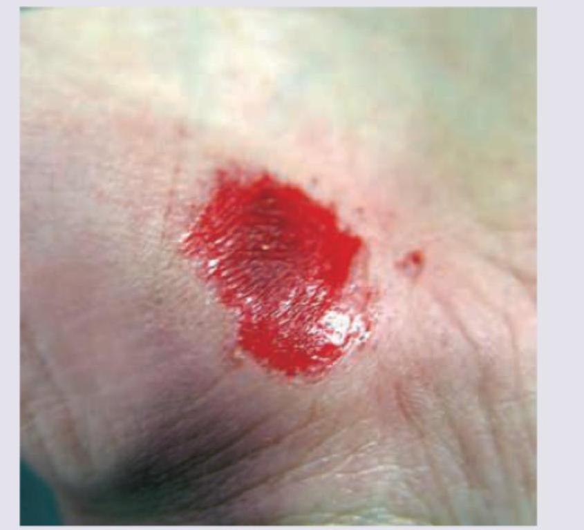
Question 2: The following image shows: (AIIMS Nov 2018)
- A. Abrasion (Correct Answer)
- B. Imprint
- C. Laceration
- D. Contusion
Explanation: ***Abrasion*** - An **abrasion** is a type of wound caused by the skin rubbing or scraping against a rough surface, resulting in the **removal of the superficial layers of the epidermis**. - The image shows a reddened, raw, and sometimes bleeding area where the top layer of skin has been scraped off, which is characteristic of an abrasion. *Imprint* - An **imprint injury** typically refers to a mark left on the skin by an object that has pressed or struck it, leaving a discernible pattern or shape. - The image does not show a distinct pattern or shape indicative of an object pressing against the skin, but rather a diffuse superficial skin loss. *Laceration* - A **laceration** is a deep cut or tear in the skin or flesh, often with irregular edges, caused by a forceful impact or trauma. - The injury in the image is superficial and does not appear to be a deep cut or tear through the skin layers. *Contusion* - A **contusion**, or bruise, is caused by trauma to blood vessels under the skin, resulting in blood leaking into surrounding tissues. - While there might be some underlying bruising, the primary visible injury is the **surface skin loss and raw appearance**, which is not characteristic of a simple contusion.
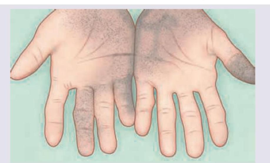
Question 3: The following presentation occurs due to? (AIIMS Nov 2018)
- A. Chronic Copper sulfate poisoning
- B. Long term ingestion of arsenic in drinking water (Correct Answer)
- C. Application of mercury ointment
- D. Application of Calotropis
Explanation: ***Long term ingestion of arsenic in drinking water*** - The image shows **hyperpigmentation and hyperkeratosis** on the palms, often described as "raindrop pigmentation" or diffuse darkening with scattered paler lesions (leukomelanosis). This is a classical dermatological manifestation of **chronic arsenic poisoning**, particularly from contaminated drinking water. - **Arsenicosis** can also cause palmar and plantar keratoses, which can progress to malignant skin lesions over time. *Chronic Copper sulfate poisoning* - Chronic copper poisoning is rare and typically affects the liver, leading to conditions like **cirrhosis** and **hepatitis**, as seen in Wilson's disease. - Skin manifestations from copper poisoning are not typically characterized by the diffuse hyperpigmentation and hyperkeratosis seen in the image. *Application of mercury ointment* - Topical mercury exposure can cause localized skin irritation, dermatitis, or systemic toxicity with features like **acrodynia** (pink disease) in children, characterized by a pink rash, irritability, and hypertension. - It does not typically lead to the described pattern of diffuse hyperpigmentation and hyperkeratosis on the palms. *Application of Calotropis* - **Calotropis** (such as *Calotropis gigantea* or *procera*) is a plant known for its toxic latex, which can cause intense local irritation, vesication, or ulceration upon skin contact. - Its effects are primarily acute irritant or allergic reactions and do not produce the chronic skin changes of hyperpigmentation and hyperkeratosis seen in the image.
Question 4: Identify the post mortem lividity of the image: (AIIMS Nov 2018)
- A. Tattooing
- B. Suggillation (Correct Answer)
- C. Putrefaction
- D. Decomposition
Explanation: ***Suggillation*** - Suggillation refers to the extravasation of blood into the tissues due to crushing pressure or trauma, often seen in cases of severe blunt force injury, leaving **diffuse, purplish discoloration.** - In post-mortem examination, a **suggillation** can be distinguished from common **livor mortis** (lividity) by its darker color and by the fact that it is not blanchable under pressure, indicating **capillary rupture and hemorrhage** rather than just pooling of blood. *Tattooing* - Tattooing involves the insertion of **indelible ink** into the dermis of the skin, creating permanent designs. - Tattoos have distinct, often patterned, appearances and would not resemble the **irregular, deep discoloration** of lividity or hemorrhage. *Putrefaction* - Putrefaction is a later stage of decomposition, characterized by the breakdown of tissues by bacteria, producing **gases, discoloration (greenish-black), and foul odors.** - This process is distinct from the **vascular pooling** or **hemorrhage** that forms suggillation; putrefaction typically starts later (after 24-48 hours) and involves more widespread tissue destruction. *Decomposition* - Decomposition is the broader process of decay of organic matter after death, encompassing various stages like **autolysis, putrefaction, and skeletonization.** - While suggillation occurs post-mortem, it is a specific type of **post-mortem bruising** or lividity, distinct from the generalized tissue breakdown and gaseous changes seen in advanced decomposition.
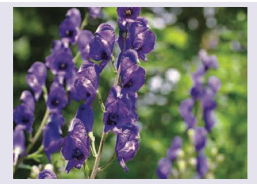
Question 5: Identify the flowers shown below:
- A. Aconite (Correct Answer)
- B. Datura
- C. Nerium Odorum
- D. Cerbera Thevetia
Explanation: ***Aconite*** - The image displays characteristics consistent with **Aconite** (monkshood) flowers, which are typically **dark purple to blue** and have a distinctive **hooded shape**. - Aconite is known for its **highly toxic** alkaloids, especially **aconitine**, which can cause rapid and severe symptoms upon ingestion or skin contact. *Cerbera Thevetia* - **Cerbera Thevetia** (yellow oleander) flowers are typically **yellow or orange** and are bell-shaped, which differs significantly from the flowers shown. - Poisoning from *Cerbera Thevetia* is primarily due to **cardiac glycosides**, leading to symptoms like bradycardia, nausea, and vomiting. *Datura* - **Datura** flowers are typically **large, trumpet-shaped**, and vary in color from white to purple, but lack the distinctive hooded appearance seen in the image. - *Datura* species contain **tropane alkaloids** (e.g., scopolamine, atropine), which cause anticholinergic effects like mydriasis, delirium, and tachycardia. *Nerium Odorum* - **Nerium Odorum** (pink oleander) flowers are typically **pink, red, or white** and have a pinwheel shape with five petals, which does not match the flowers in the image. - Like *Thevetia*, *Nerium Odorum* is toxic due to **cardiac glycosides**, causing similar cardiovascular and gastrointestinal symptoms.
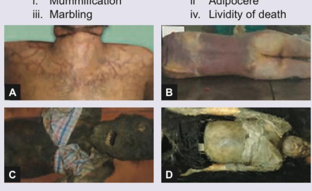
Question 6: Identify the correct pair: i. Mummification ii. Adipocere iii. Marbling iv. Livor mortis
- A. i-D, ii-C, iii-B, iv-A (Correct Answer)
- B. i-C, ii-D, iii-B, iv-A
- C. i-C, ii-D, iii-A, iv-B
- D. i-D, ii-A, iii-C, iv-B
Explanation: ***i-D, ii-C, iii-B, iv-A*** - Image **D** clearly shows a body exhibiting **mummification**, characterized by a dried, leathery appearance due to dehydration in dry conditions. - Image **C** depicts **adipocere**, where fatty tissues are converted into a grayish-white, greasy, or waxy substance, which can preserve body contours. - Image **B** illustrates **marbling**, which is a pattern of greenish-black discoloration along superficial veins due to the breakdown of blood by putrefactive bacteria. - Image **A** displays **lividity of death (livor mortis)**, characterized by purplish discoloration of the skin in dependent areas due to the gravitational pooling of blood after circulation ceases.
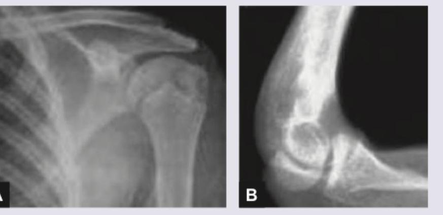
Question 7: Comment on age of the male patient:
- A. 5-7 years
- B. 12-14 years (Correct Answer)
- C. 17-19 years
- D. >25 years
Explanation: ***12-14 years*** - The radiographs show **open growth plates** (epiphyseal lines) in both the shoulder (A) and elbow (B), indicating the individual is still growing. - The presence of well-developed secondary ossification centers and significant but not fully fused physes is consistent with a male in the **mid-pubertal** range, typically observed between 12-14 years. *5-7 years* - At this age, many **secondary ossification centers** would just be appearing or still quite small, and the growth plates would be much wider and less defined than seen in the images. - The degree of skeletal maturation evident in the shoulder and elbow in the images surpasses that of a 5-7 year old. *17-19 years* - By this age, most **growth plates** in males, especially in key joints like the shoulder and elbow, would be largely **fused** or in the final stages of fusion. - The distinct open physes seen in both images rule out this age range, as significant growth is still occurring. *>25 years* - In an individual over 25 years, **all growth plates** would be completely **fused**, and there would be no visible epiphyseal lines. - The presence of clear, open growth plates in the images definitively excludes an adult age.