INI-CET 2018
68 Previous Year Questions with Answers & Explanations
Anatomy
2 questionsCranial nerve that is involved in olfaction:
Identify the structure marked in the image of cardiac myocyte. (AIIMS Nov 2018)
INI-CET 2018 - Anatomy INI-CET Practice Questions and MCQs
Question 1: Cranial nerve that is involved in olfaction:
- A. Hypoglossal
- B. Trigeminal (Correct Answer)
- C. Glossopharyngeal
- D. Vagus
Explanation: ***Trigeminal*** - The **trigeminal nerve (CN V)** carries sensory information from the face and contributes to olfactory perception through its **ophthalmic (V1) and maxillary (V2) divisions** [1]. - These divisions detect **chemical irritants, pungency, cooling, and warming sensations** in the nasal cavity (chemesthesis), contributing to the overall perception of smells. - Examples include the stinging sensation of **ammonia**, cooling of **menthol**, and burning of **capsaicin** - all part of the smell experience. - This is distinct from true olfaction (CN I) but is an essential component of how we perceive "smell" in daily life. *Hypoglossal* - The **hypoglossal nerve (CN XII)** is purely a **motor nerve** that controls the intrinsic and extrinsic muscles of the **tongue**. - It is essential for **speech, swallowing, and tongue movement** but has **no role in olfaction or smell perception**. *Glossopharyngeal* - The **glossopharyngeal nerve (CN IX)** mediates **taste sensation from the posterior third of the tongue**, swallowing, and salivation. - While important for taste, it has **no role in olfactory pathways or smell detection**. *Vagus* - The **vagus nerve (CN X)** has extensive **parasympathetic functions** and carries taste sensation from the **epiglottis and pharynx**. - It innervates thoracic and abdominal organs but is **not involved in olfactory perception**.
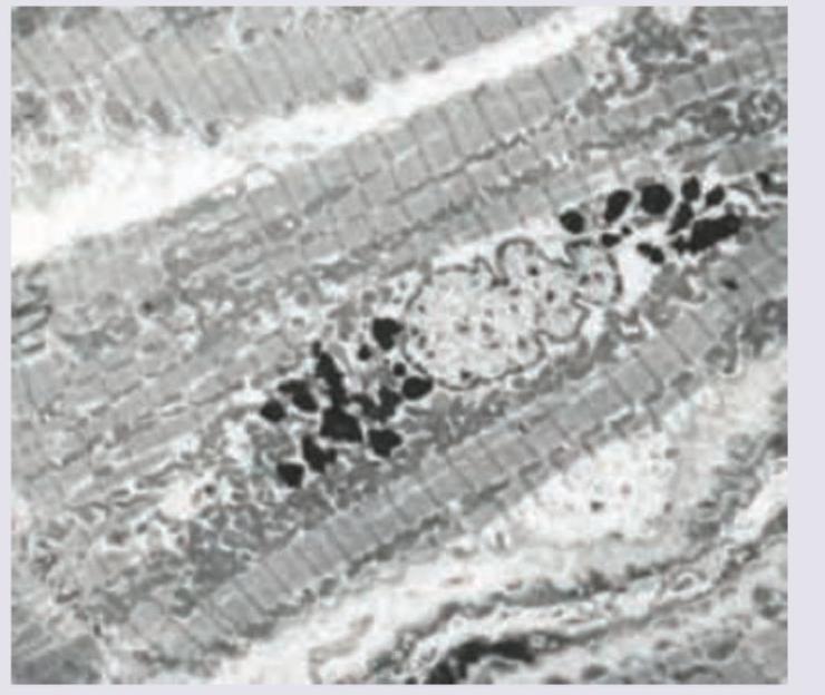
Question 2: Identify the structure marked in the image of cardiac myocyte. (AIIMS Nov 2018)
- A. Lipofuscin granules (Correct Answer)
- B. Lysosomes
- C. Phagolysosome
- D. Inflammasome
Explanation: ***Lipofuscin granules*** - The image displays electron-dense, irregular structures, characteristic of **lipofuscin granules**, which are wear-and-tear pigments accumulating in aging cardiac myocytes. - They are typically located near the nucleus and appear dark due to their complex lipid-protein composition. *Lysosomes* - Lysosomes are typically smaller, more uniformly shaped, and contain hydrolytic enzymes, which is not clearly depicted. - While involved in cellular waste breakdown, they do not typically accumulate as large, intensely electron-dense, irregular aggregates like those shown. *Phagolysosome* - A phagolysosome forms when a phagosome fuses with a lysosome, containing engulfed material often of foreign or cellular debris origin. - The image does not show evidence of recently engulfed material or the typical morphology of a phagocytic vesicle. *Inflammasome* - An inflammasome is a multi-protein intracellular complex involved in the inflammatory response, not a visibly distinct organelle with this characteristic electron microscopic appearance. - It is a signaling platform, not a storage granule, and would not appear as dense, granular deposits in a routine electron micrograph.
Pathology
3 questionsWhich of the following tubes contain sodium fluoride as an anti-coagulant?
A patient presents with intermittent fever, no weight loss and enlarged retroperitoneal lymph nodes. Peripheral smear is normal. Gross sample and its histopathology slide is shown below. Comment on the diagnosis.
Immunofluorescence staining pattern from a kidney biopsy from a 35-year-old patient presenting with proteinuria has been shown below. What is the most probable cause? (AIIMS Nov 2018)
INI-CET 2018 - Pathology INI-CET Practice Questions and MCQs
Question 1: Which of the following tubes contain sodium fluoride as an anti-coagulant?
- A. Gray-top tube (Correct Answer)
- B. Green-top tube
- C. Pink-top tube
- D. Blue-top tube with yellow checkerboard pattern
Explanation: ***a. Gray-top tube*** - The **gray-top tube** contains **sodium fluoride** and **potassium oxalate** as anticoagulants. - Sodium fluoride acts as both an **anticoagulant** and an **antiglycolytic agent**, preserving glucose concentration by inhibiting glycolysis. - This tube is the **standard choice for glucose testing** in clinical laboratories. *b. Green-top tube* - The **green-top tubes** contain **heparin** (either sodium, lithium, or ammonium heparin) as an anticoagulant. - These tubes are used for various tests including **plasma chemistry** and some hormone assays, but not for glucose preservation with sodium fluoride. *c. Pink-top tube* - The **pink-top tube** generally contains **EDTA** (ethylenediaminetetraacetic acid) and is specifically designed for **blood bank** tests due to its special labeling and anticoagulant properties for cross-matching. - While it contains an anticoagulant, it is not sodium fluoride and is not used for glucose measurement. *d. Blue-top tube with yellow checkerboard pattern* - While some manufacturers may use different labeling systems, the **standard tube for sodium fluoride** is the gray-top tube, not the blue-top with checkerboard pattern. - Blue-top tubes typically contain **sodium citrate** and are used for coagulation studies (PT, PTT, INR).
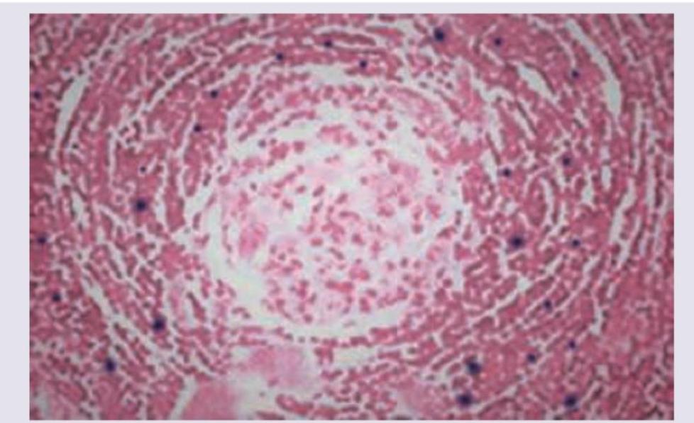
Question 2: A patient presents with intermittent fever, no weight loss and enlarged retroperitoneal lymph nodes. Peripheral smear is normal. Gross sample and its histopathology slide is shown below. Comment on the diagnosis.
- A. Non-Hodgkin's lymphoma
- B. Castleman disease (Correct Answer)
- C. Angiolymphoid hyperplasia
- D. IgG4 related disease
Explanation: ***Castleman disease*** - The image shows **"lollipop lesions"** or **"onion-skinning"** of follicles with **regressed germinal centers** and prominent mantle zones, characteristic of the hyaline-vascular variant of Castleman disease. - Clinical features like **intermittent fever** and **enlarged retroperitoneal lymph nodes** with **no weight loss** can be seen in Castleman disease, particularly the unicentric type. *Non-Hodgkin's lymphoma* - This would typically show effacement of the normal lymph node architecture by a **monotonous proliferation of atypical lymphocytes**. - While it can cause lymphadenopathy and fever, the specific histological features presented do not align with typical non-Hodgkin's lymphoma. *Angiolymphoid hyperplasia* - Characterized by **multiple vascular channels** lined by plump endothelial cells with **lymphoid infiltrates**. - It often presents as subcutaneous nodules, usually in the head and neck region, and lacks the follicular changes seen here. *IgG4 related disease* - Histologically, this disease is characterized by a **dense lymphoplasmacytic infiltrate rich in IgG4-positive plasma cells**, storiform fibrosis, and obliterative phlebitis. - While it can cause lymph node enlargement, the specific follicular changes in the image are not typical of IgG4-related disease.
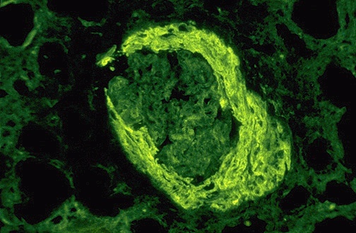
Question 3: Immunofluorescence staining pattern from a kidney biopsy from a 35-year-old patient presenting with proteinuria has been shown below. What is the most probable cause? (AIIMS Nov 2018)
- A. FSGS
- B. PSGN
- C. Lupus nephritis (Correct Answer)
- D. Goodpasture syndrome
- E. IgA nephropathy
Explanation: ***Lupus nephritis*** - The image likely shows a **"full house" immunofluorescence pattern** (deposits of IgG, IgA, IgM, C3, and C1q), which is characteristic of **lupus nephritis** [1]. - This pattern indicates immune complex deposition in the glomeruli, a hallmark of systemic lupus erythematosus affecting the kidneys [1]. *FSGS* - **Focal segmental glomerulosclerosis (FSGS)** typically shows **negative immunofluorescence** or only non-specific IgM and C3 in sclerotic areas [3,4]. - It does not present with the diffuse, multi-immunoglobulin deposition seen in the characteristic "full house" pattern. *PSGN* - **Post-streptococcal glomerulonephritis (PSGN)** characteristically shows a **"starry sky" or granular pattern** of C3 and IgG deposition, often with a dominant C3 [2]. - It does not typically show the full spectrum of immunoglobulins and complement components seen in lupus nephritis. *Goodpasture syndrome* - **Goodpasture syndrome** is characterized by a **linear deposition of IgG** along the glomerular basement membrane (GBM) on immunofluorescence [2]. - This pattern is distinct from the granular, multi-immunoglobulin deposition seen in immune complex-mediated diseases like lupus nephritis. **References:** [1] Cross SS. Underwood's Pathology: A Clinical Approach. 6th ed. Common Clinical Problems From Diseases Of The Urinary And Male Genital Tracts, pp. 532-533. [2] Kumar V, Abbas AK, et al.. Robbins and Cotran Pathologic Basis of Disease. 9th ed. The Kidney, p. 915. [3] Cross SS. Underwood's Pathology: A Clinical Approach. 6th ed. Common Clinical Problems From Diseases Of The Urinary And Male Genital Tracts, pp. 530-531. [4] Cross SS. Underwood's Pathology: A Clinical Approach. 6th ed. Common Clinical Problems From Diseases Of The Urinary And Male Genital Tracts, pp. 531-532.
Physiology
2 questionsA macrophage engulfs different cells as shown in the image. This is known as?
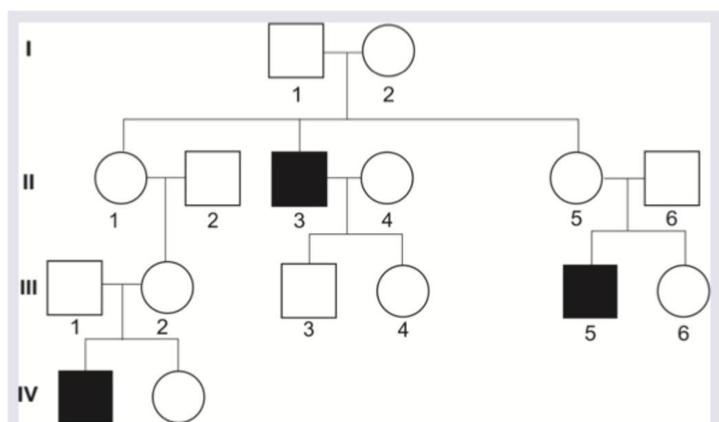
Identify the inheritance pattern shown below.
INI-CET 2018 - Physiology INI-CET Practice Questions and MCQs
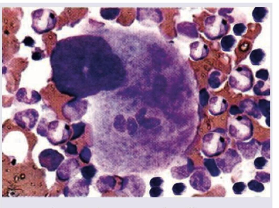
Question 1: A macrophage engulfs different cells as shown in the image. This is known as?
- A. Phagocytosis
- B. Killing
- C. Cytotoxicity
- D. Emperipolesis (Correct Answer)
Explanation: ***Emperipolesis*** - The image shows a large cell (likely a macrophage or megakaryocyte) containing **intact various blood cells** within its cytoplasm without signs of degeneration. - **Emperipolesis** is specifically defined as the **active penetration of one cell by another**, where both the engulfed and engulfing cells remain viable. *Phagocytosis* - **Phagocytosis** involves the ingestion and subsequent **destruction or degradation** of foreign particles, microorganisms, or cellular debris. - The cells within the macrophage in the image appear **morphologically intact** and not in a state of degradation. *Killing* - **Killing** implies the process by which a cell actively destroys another cell, often through mechanisms like **apoptosis or necrosis**. - There are **no morphological features** in the image to suggest that the engulfed cells are being actively killed or are undergoing degeneration. *Cytotoxicity* - **Cytotoxicity** refers to the ability of certain immune cells (e.g., cytotoxic T lymphocytes, NK cells) to **kill target cells**. - This process usually involves specific recognition and induction of target cell death, which is not what is depicted by the presence of intact cells within another cell.
Question 2: Identify the inheritance pattern shown below.
- A. Y linked recessive
- B. X linked recessive (Correct Answer)
- C. X linked dominant
- D. Y linked dominant
Explanation: ***X linked recessive*** - The pattern shows that mainly **males are affected**, and the trait skips generations (e.g., individual I-1 is unaffected, but his children are affected). - Affected fathers (like II-3) do not pass the trait to their sons, but their daughters are carriers and can pass it on to their sons (like IV-1). *Y linked recessive* - In Y-linked inheritance, only **males would be affected**, and all sons of an affected father would inherit the trait. - This pedigree shows unaffected fathers having affected sons, and not all sons of affected males are affected directly. *X linked dominant* - In X-linked dominant inheritance, affected fathers pass the trait to **all their daughters**, and at least one parent would be affected in each generation. - This pedigree shows skipping of generations and affected individuals being born to unaffected parents (e.g., II-3 and III-5). *Y linked dominant* - Y-linked inheritance, whether dominant or recessive, would only affect **males** and would be directly passed from father to all sons. - The pedigree shows unaffected parents having affected offspring (e.g., I-1 and I-2 produced II-3), which rules out Y-linked inheritance.
Radiology
3 questionsWhat does the given chest X-ray show?
Identify the type of investigation shown in the image below.
A 25-year-old female presents with neck pain and tingling sensation in her left arm. An X-ray of the cervicothoracic region is obtained. What is the radiological finding shown in the image?
INI-CET 2018 - Radiology INI-CET Practice Questions and MCQs
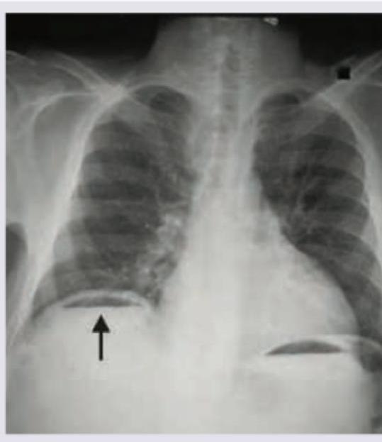
Question 1: What does the given chest X-ray show?
- A. Pneumoperitoneum (Correct Answer)
- B. Emphysema
- C. Diaphragmatic hernia
- D. Diaphragmatic eventration
Explanation: ***Pneumoperitoneum*** - The chest X-ray shows **free air under the diaphragm**, visible as a lucent (dark) crescent between the liver/spleen and the diaphragm (indicated by the arrow on the right side of the patient). - This finding is diagnostic of **pneumoperitoneum**, which is often caused by a perforated abdominal viscus like a peptic ulcer or bowel perforation. *Emphysema* - **Emphysema** is a lung condition characterized by over-inflated alveoli and air trapping within the lungs, leading to hyperlucency of the lung fields and flattened diaphragms. - It does not present as free air below the diaphragm but rather as changes within the lung parenchyma. *Diaphragmatic hernia* - A **diaphragmatic hernia** involves the protrusion of abdominal organs into the chest cavity through a defect in the diaphragm. - This would typically show abdominal contents (e.g., bowel loops or stomach) above the diaphragm in the thoracic cavity, not free air below it. *Diaphragmatic eventration* - **Diaphragmatic eventration** is an abnormal elevation of part or all of an intact hemidiaphragm due to thinning and weakness of the diaphragmatic muscle. - It causes an elevated diaphragm but does not involve free air in the peritoneal cavity.
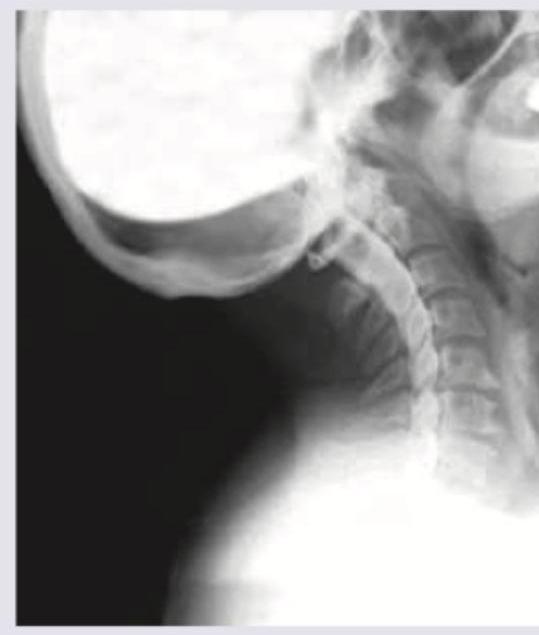
Question 2: Identify the type of investigation shown in the image below.
- A. Angiography
- B. Myelography (Correct Answer)
- C. Neurography
- D. Fluoroscopy
Explanation: ***Correct: Myelography*** - The image displays **contrast agent within the spinal canal**, outlining the spinal cord and nerve roots against the bony structures of the cervical spine - This technique is used to visualize **nerve impingement, disc herniation, or spinal cord compression** - Characteristic finding: contrast delineating the thecal sac and nerve root sleeves *Incorrect: Angiography* - Angiography involves injecting contrast into **blood vessels** to visualize vascular structures, detect blockages, or aneurysms - The image shows the **spinal canal** rather than the vascular tree *Incorrect: Neurography* - Neurography (MR neurography) is a specialized **MRI technique** to visualize peripheral nerves themselves - Does not involve injection of contrast into the spinal canal as shown in the image *Incorrect: Fluoroscopy* - Fluoroscopy is a **real-time X-ray imaging technique** used for dynamic assessment or procedure guidance - While fluoroscopy may be used **during** myelography to guide needle placement, the specific technique of contrast visualization in the spinal canal defines this as myelography
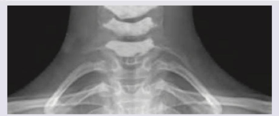
Question 3: A 25-year-old female presents with neck pain and tingling sensation in her left arm. An X-ray of the cervicothoracic region is obtained. What is the radiological finding shown in the image?
- A. Cervical rib (Correct Answer)
- B. Costochondritis
- C. Fracture of 2nd rib
- D. Spondylolisthesis
Explanation: ***Cervical rib*** - The image displays an extra rib arising from the **C7 cervical vertebra**, which is characteristic of a cervical rib. - This **supernumerary rib** extends towards the sternum or first thoracic rib, a classic radiological finding. *Costochondritis* - **Costochondritis** is an inflammation of the cartilage connecting the ribs to the sternum, which is typically a clinical diagnosis, not visible on X-ray. - An X-ray would not show inflammatory changes in cartilage or soft tissue, making this diagnosis unlikely based on imaging alone. *Fracture of 2nd rib* - A **fracture of the 2nd rib** would appear as a discontinuity or break in the normal bony architecture of the second rib. - The image does not show any signs of a broken rib; instead, it shows an **extra, well-formed rib-like structure** originating from the cervical spine. *Spondylolisthesis* - **Spondylolisthesis** involves the anterior displacement of one vertebral body over another, usually in the lumbar spine. - This condition is also not visible in the provided image, which focuses on the cervicothoracic junction and shows an **anatomic variation** rather than vertebral slippage.
About INI-CET 2018 Questions
This page contains 68 questions from the INI-CET 2018 paper, organised across 15 subjects for focused practice. Every question comes with the correct answer and a detailed explanation to help you understand the underlying concept. Subject-wise organisation lets you target specific areas and identify which topics carried the most weight in this particular year.
Practising year-wise papers is essential for understanding how the INI-CET exam evolves — you can spot trending topics, gauge difficulty shifts, and benchmark your readiness against a real paper. To take your preparation further, download the Oncourse app for AI-driven performance insights, spaced repetition of questions you got wrong, and a personalised study plan built around your INI-CET goals.