
Abdominal and Pelvic Radiology — Flashcards

On this page
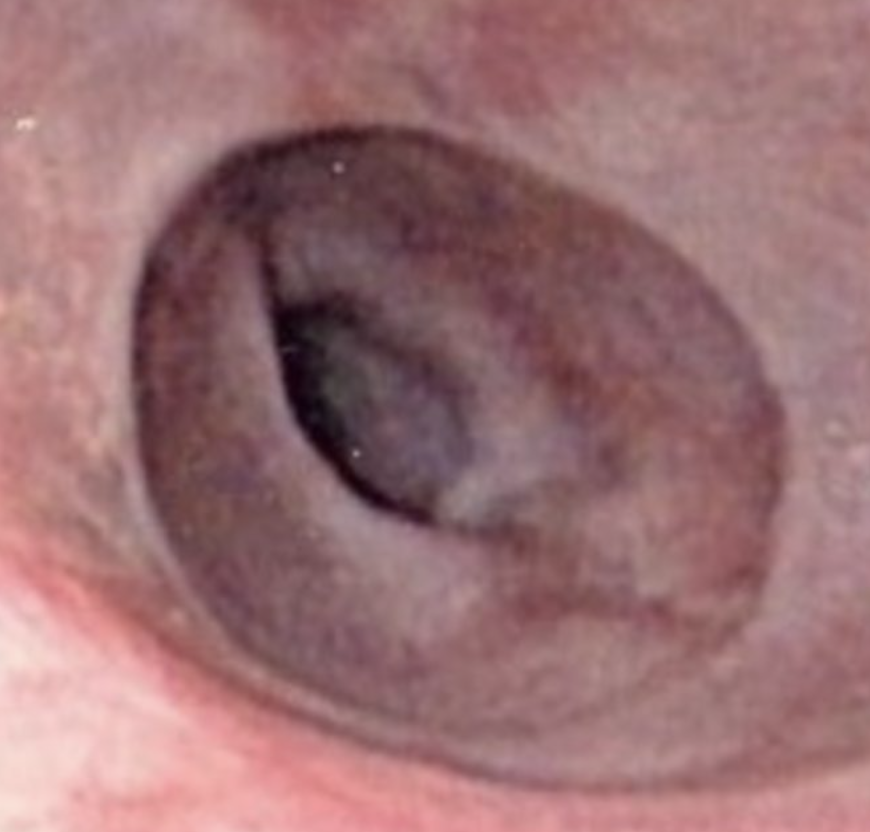
The given image shows the presence of a _____ in lower end of esophagus.
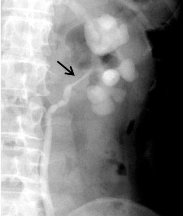
The given Retrograde ureterogram showing dilated clubbed calyces with stricture of the left ureteropelvic junction is suggestive of _____
In _____ sign or double wall sign, inner mucosal and outer serosal layers of bowel are enhanced in pneumoperitoneum
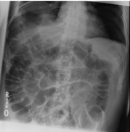
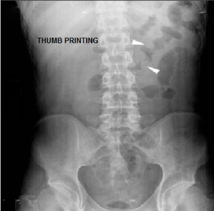
Thumbprinting sign is suggestive of _____
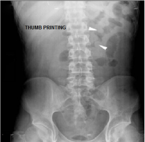
The thumbprinting sign is seen in both _____ colitis and _____ colitis.
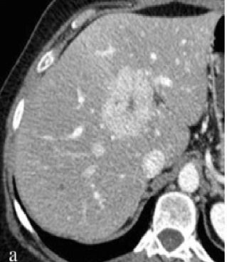
Hypervascular liver lesion with a central non-enhancing scar points to the diagnosis of _____
Bilateral _____ ureters describes the appearance of the distal ureter in patients with significant benign prostatic hypertrophy.
_____ deformity is the appearance of the deviation of bilateral ureters seen classically in retroperitoneal fibrosis
_____ sign on CT is seen in Midgut volvulus
The _____ sign (or champagne glass sign) refers to the appearance of the ureter when it is focally dilated by an intraluminal mass and is best seen by a retrograde ureterogram.
Study by Chapter
Imaging of Liver
Flashcards
Biliary Tract Imaging
Flashcards
Pancreatic Imaging
Flashcards
Spleen and Lymphatic System
Flashcards
Gastrointestinal Tract Imaging
Flashcards
Renal and Urinary Tract Imaging
Flashcards
Adrenal Imaging
Flashcards
Female Pelvic Imaging
Flashcards
Male Pelvic Imaging
Flashcards
Abdominal Trauma Imaging
Flashcards
Acute Abdomen Imaging
Flashcards
Imaging of Peritoneal Cavity and Retroperitoneum
Flashcards
About Abdominal and Pelvic Radiology Flashcards for NEET-PG
These Abdominal and Pelvic Radiology flashcards are designed for NEET-PG Radiology preparation, using active recall to help you retain high-yield concepts, clinical correlations, and commonly tested facts. Each card prompts you to retrieve information from memory rather than passively reviewing notes, which research shows leads to significantly better exam performance.
The 129 cards in this deck cover the most important topics in Abdominal and Pelvic Radiology, including key mechanisms, diagnostic criteria, treatment protocols, and clinical pearls that frequently appear in NEET-PG examinations. Cards are organised by chapter so you can focus on specific areas or work through the entire topic systematically.
For personalised spaced repetition scheduling that adapts to your performance, unlimited flashcards, and detailed progress analytics, download the Oncourse app.
Frequently Asked Questions
Are Abdominal and Pelvic Radiology flashcards free on Oncourse?
How do flashcards help with NEET-PG preparation?
How many Abdominal and Pelvic Radiology flashcards are available?
Can I study Abdominal and Pelvic Radiology flashcards by chapter?
What topics do these Radiology flashcards cover?
Want unlimited flashcards?
Get full access to all flashcards, spaced repetition, and progress tracking.
Start For Free