
Imaging of Liver — Flashcards

HCCs often appears _____vascular during the arterial phase of computed tomography (CT) studies
Hint: hypo/hyper
_____ sign: enhancing dots within the dilated intrahepatic bile ducts, representing portal radicles is seen on CT in Caroli's disease
_____ in case of liver hemangioma shows Light bulb sign
_____ phase CT demonstrates hypervascularity with a central stellate scar, in the case of Focal nodular hyperplasia
_____ is the investigation of choice in a liver hemangioma
How often should you perform imaging follow-up for an inflammatory hepatocellular adenoma (IHCA)?
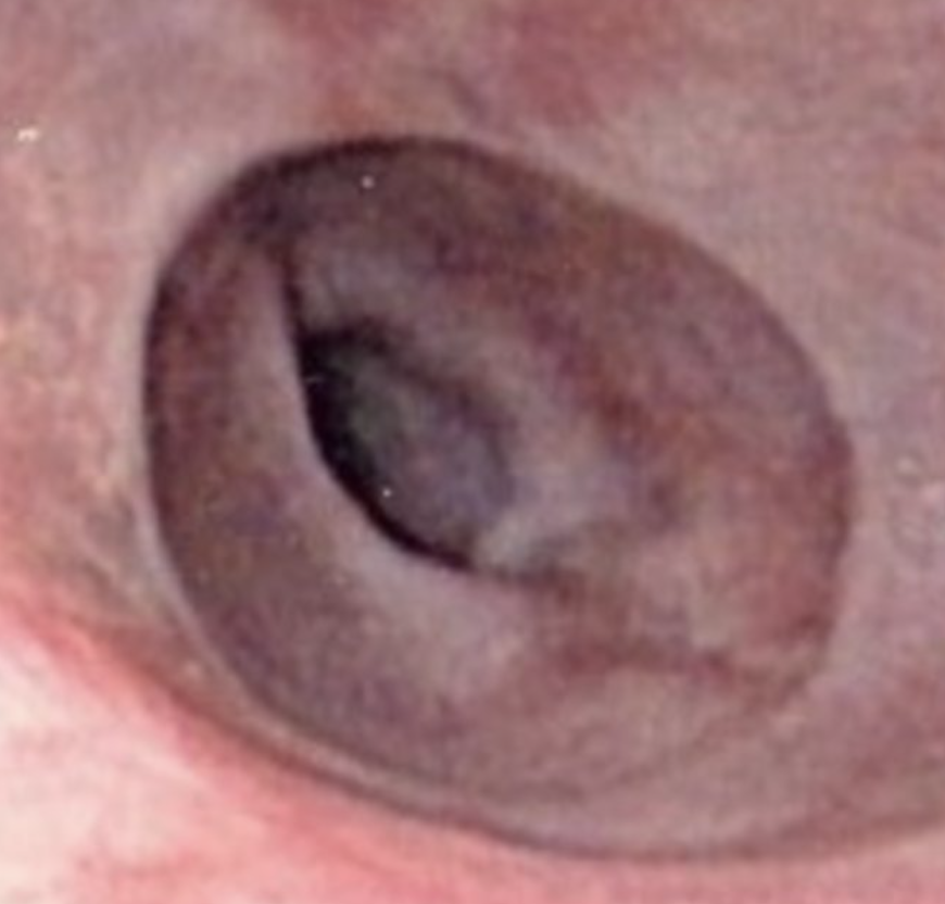
The given image shows the presence of a _____ in lower end of esophagus.
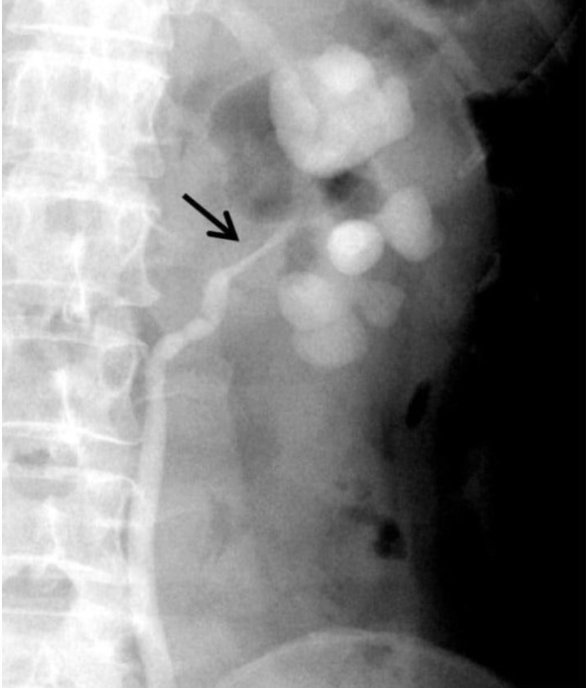
The given Retrograde ureterogram showing dilated clubbed calyces with stricture of the left ureteropelvic junction is suggestive of _____
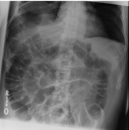
In _____ sign or double wall sign, inner mucosal and outer serosal layers of bowel are enhanced in pneumoperitoneum
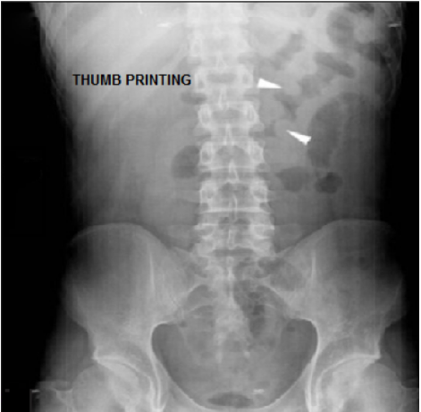
Thumbprinting sign is suggestive of _____
Imaging of Liver Flashcards for NEET-PG
Study 10 flashcards on Imaging of Liver for NEET-PG Radiology. These active recall cards cover the key concepts, clinical associations, and high-yield facts from this chapter of Abdominal and Pelvic Radiology. Each card is designed to test your understanding rather than just recognition, building stronger and more durable memories for exam day.
For personalised spaced repetition scheduling and unlimited flashcards, download the Oncourse app.
Frequently Asked Questions
Are Imaging of Liver flashcards free?
How many flashcards does this chapter have?
How should I use these flashcards for NEET-PG?
Are there more flashcards for Abdominal and Pelvic Radiology?
Want unlimited flashcards?
Get full access to all flashcards, spaced repetition, and progress tracking.
Scan to download app