
Reproductive Pathology — Flashcards

On this page
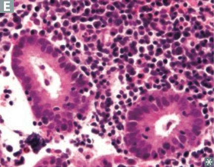
The histology image below has lymphocytes and plasma cells in the endometrium, which is indicative of _____
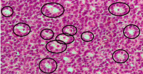
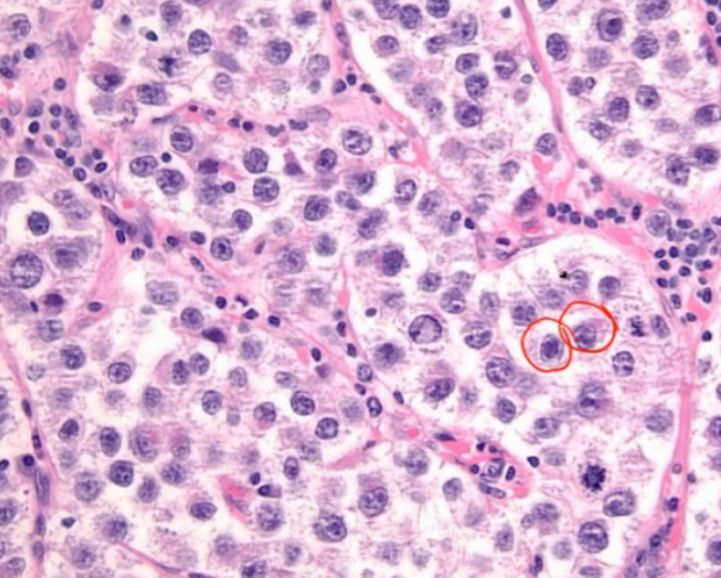
The image shows _____ bodies which are seen in Granulosa cell tumour.
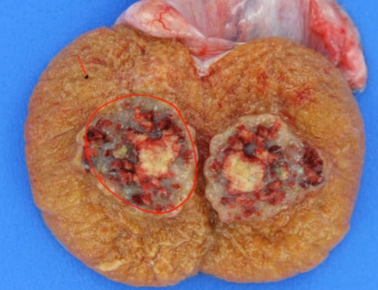
_____ is a testicular germ cell tumor that forms a hemorrhagic mass with necrosis
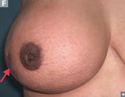
The gross appearance of inflammatory breast cancer is often described as resembling an _____ (due to invasion of lymphatic spaces)
_____ is a malignant testicular tumor comprised of large cells with clear cytoplasm and central nuclei, sometimes described as a "fried egg" appearance
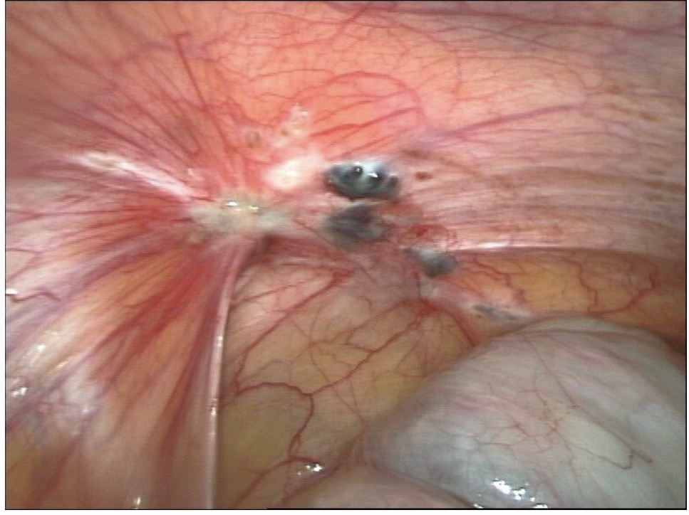
Powder burn spots over the peritoneum are seen in _____.
Small _____philic colloid amyloid bodies called corpora amylacea are frequently seen within a prostatic follicle.
_____ emboli are characterized by squamous cells and keratin debris in the embolus
_____ are the most common changes in pre-menopausal breast
The Gleason grading system (for prostatic adenocarcinoma) is based on _____, not nuclear atypia
Study by Chapter
Diseases of Male Genital Tract
Flashcards
Testicular Tumors
Flashcards
Prostate Pathology
Flashcards
Diseases of Female Genital Tract
Flashcards
Cervical Pathology and Neoplasia
Flashcards
Endometrial Pathology
Flashcards
Ovarian Diseases and Tumors
Flashcards
Gestational Trophoblastic Disease
Flashcards
Placental Pathology
Flashcards
Sexually Transmitted Infections
Flashcards
Want unlimited flashcards?
Get full access to all flashcards, spaced repetition, and progress tracking.
Scan to download app