Anatomy
1 questionsWhich of the following is a remnant of the Wolffian duct in females?
Community Medicine
1 questionsThe difference between the incidence in the exposed and non-exposed group is best given by:
Microbiology
3 questionsThe image of an immunoglobulin is shown below. Which type of immunoglobulin is it?

Which of the following does not possess superantigen properties?
Which antibody is not transmitted from mother to baby?
Obstetrics and Gynecology
1 questionsWhat is the use of the instrument shown in the image?

Pathology
1 questionsA 33-year-old man presents with a 5-week history of calf pain, swelling, and low-grade fever. Serum levels of creatinine kinase are elevated. A muscle biopsy reveals numerous eosinophils and he also has peripheral blood eosinophilia. Which of the following interleukins is primarily responsible for the increase in eosinophils in this patient?
Pediatrics
1 questionsWhich condition is characterized by conjunctival injection, pharyngeal injection, polymorphic rash, and cervical lymphadenopathy?
Physiology
1 questionsA 35-year-old female experiences a tingling sensation in her arm after watching TV for long hours with her hands under her head. Which type of nerve fibers is most likely to be affected due to this position?
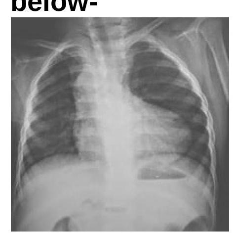
Radiology
1 questionsIdentify the condition in the X-ray given below: