All SubjectsAnatomy (110)Anesthesiology (34)Biochemistry (129)Community Medicine (109)Dental (16)Dermatology (34)ENT (62)Forensic Medicine (100)General Medicine (2)Internal Medicine (120)Microbiology (108)Obstetrics and Gynecology (79)Ophthalmology (78)Orthopaedics (41)Pathology (90)Pediatrics (33)Pharmacology (134)Physiology (91)Psychiatry (6)Psychiatry (81)Radiology (41)Surgery (52)
Q61
Milk ring test is done to detect which organism present in milk?
Q62
Prions are best killed by
Q63
Which is not a DNA virus?
Q64
Which of the following is a superantigen ?
Q65
What are the changes in the variable region of immunoglobulins?
Q66
Which of the following bacteria is not capsulated?
Q67
What is the cause of rabies in wild animals?
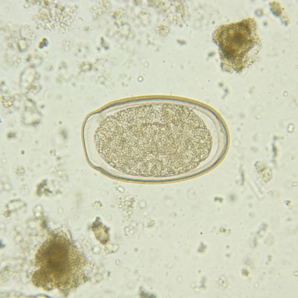
Q68
Child having perianal pruritus with the following eggs is due to -
Q69
The fungus with septate hyphae and dichotomous branching is?
Q70
Which of the following statements about Helminths is false?