
General Microbiology — MCQs

On this page
What is the approximate ratio of anaerobes to aerobes found in normal human stool?
Which of the following is a prokaryote?
Microbiological waste should be segregated in which color bag?
Which of the following is NOT a function of flagella?
Endotoxin consists of which of the following components?
Some microorganisms produce a diffuse spreading inflammatory reaction due to the elaboration of which enzyme?
The toughness of bacterial cell walls is due to:
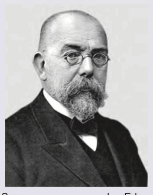
Identify the scientist shown in the image below:
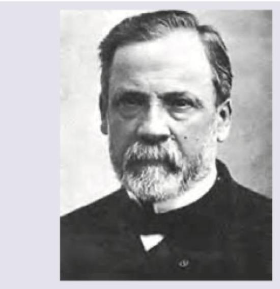
Identify the scientist given in the image below:
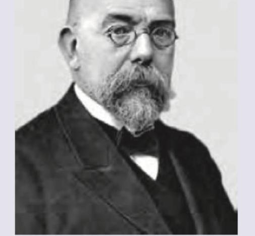
Identify the scientist. (Recent NEET Pattern 2016-17)
Practice by Chapter
History and Scope of Microbiology
Practice Questions
Classification of Microorganisms
Practice Questions
Bacterial Morphology and Structure
Practice Questions
Bacterial Physiology and Metabolism
Practice Questions
Bacterial Genetics
Practice Questions
Microbial Growth and Nutrition
Practice Questions
Sterilization and Disinfection
Practice Questions
Bacterial Identification Methods
Practice Questions
Normal Microbiota and Pathogenicity
Practice Questions
Antimicrobial Susceptibility Testing
Practice Questions
Want unlimited practice?
Get full access to all questions, explanations, and performance tracking.
Scan to download app