
Diagnostic Microbiology — MCQs

On this page
The Widal test is a type of:
Heating and subsequent plating is a method used for isolating which of the following organisms?
What dye is used in fluorescent microscopy?
Which of the following is an acid-fast organism?
Which of the following serological tests is NOT helpful in the diagnosis of chronic brucellosis?
The Quellung reaction is characteristic of which bacterium?
Streptococcal glomerulonephritis is best diagnosed by:
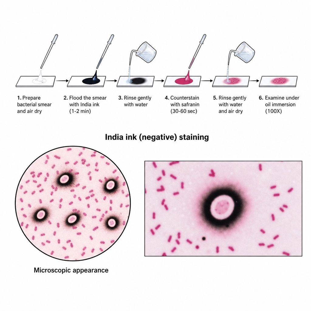
The staining procedure shown in the illustration is used to visualize which of the following structures?
A patient presents with a 14-day history of fever, and typhoid fever is suspected. Which investigation is most appropriate for diagnosis?
A 50-year-old male presented with non-destructive migratory arthritis of the left lower limb joints, intermittent fever, myalgias, multiple episodes of diarrhea, abdominal pain, and significant weight loss. Examination revealed cervical lymphadenopathy, hepatosplenomegaly, skin hyperpigmentation, and mild anterior uveitis. Laboratory findings included negative rheumatoid factor and ANA, anemia, and hypereosinophilia. Endoscopy showed pale, yellow, shaggy mucosa with erythema and ulceration past the first part of the duodenum. Biopsy of the small intestine was taken, and PAS staining was performed. PCR was ordered to diagnose the condition. Which RNA sequencing is used to diagnose the above condition?
Practice by Chapter
Specimen Collection and Transport
Practice Questions
Microscopy in Microbiology
Practice Questions
Culture Methods and Media
Practice Questions
Bacterial Identification Techniques
Practice Questions
Antimicrobial Susceptibility Testing
Practice Questions
Serological Diagnosis
Practice Questions
Molecular Diagnostic Methods
Practice Questions
Rapid Diagnostic Tests
Practice Questions
Point-of-Care Testing
Practice Questions
Automation in Microbiology Laboratory
Practice Questions
Quality Control in Diagnostic Microbiology
Practice Questions
Interpretation of Microbiological Reports
Practice Questions
Want unlimited practice?
Get full access to all questions, explanations, and performance tracking.
Scan to download app