
Diagnostic Microbiology — MCQs

On this page
Infection with Epstein-Barr virus (EBV) results in the development of virus-specific antibodies. The pattern of these antibodies helps to stage the illness. Which statement regarding EBV-VCA (IgG) Ab is correct?
Corynebacterium diphtheriae produces black or grey colonies on which of the following agar media?
A 24-year-old cook from a hostel mess suffered from enteric fever two years ago. How can the chronic carrier state in this patient be diagnosed?
Under the Revised National Tuberculosis Control Programme (RNTCP), which method is used for drug susceptibility testing?
Modified Thayer-Martin medium is used for the isolation of which bacterium?
A young male is admitted with a history of altered sensorium and hydrophobia. A clinical diagnosis of rabies was made, and corneal scrapings were taken. Which is the best test to confirm his diagnosis?
A false negative tuberculin reaction may be obtained in all of the following situations except?
What is MYPA agar used for?
What is the standard screening test for syphilis?
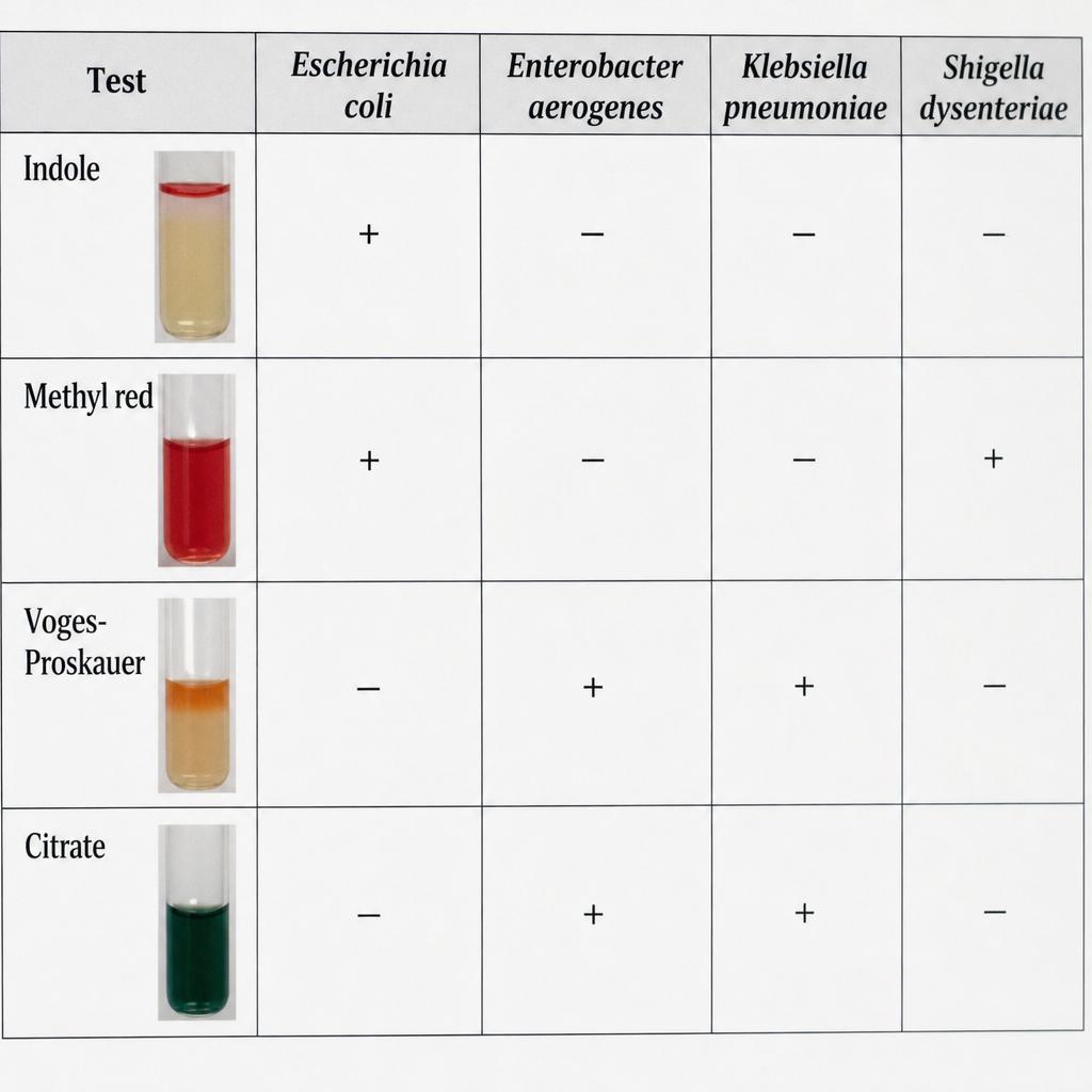
Which bacteria are indicated by this IMViC pattern of biochemical reactions?
Practice by Chapter
Specimen Collection and Transport
Practice Questions
Microscopy in Microbiology
Practice Questions
Culture Methods and Media
Practice Questions
Bacterial Identification Techniques
Practice Questions
Antimicrobial Susceptibility Testing
Practice Questions
Serological Diagnosis
Practice Questions
Molecular Diagnostic Methods
Practice Questions
Rapid Diagnostic Tests
Practice Questions
Point-of-Care Testing
Practice Questions
Automation in Microbiology Laboratory
Practice Questions
Quality Control in Diagnostic Microbiology
Practice Questions
Interpretation of Microbiological Reports
Practice Questions
Want unlimited practice?
Get full access to all questions, explanations, and performance tracking.
Scan to download app