
Bacteriology — MCQs

On this page
Which of the following statements about E. coli are true?
A patient presents with fever for 3 weeks. On examination, he is observed to have splenomegaly. Ultrasonography reveals a hypoechoic shadow in the spleen near the hilum. Gram-negative bacilli are isolated on blood culture. Which of the following is the most likely causative organism?
The appearance of cobweb formation in CSF indicates which of the following?
Mycoplasma organisms may also cause disease in non-pulmonary sites. Which of the following is the most commonly affected non-pulmonary site?
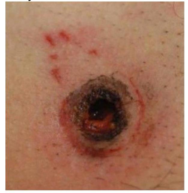
What is the likely cause of this manifestation?
Which of the following is a tripartite toxin?
Which of the following is true about Clostridium tetani?
What is true about Protein A of Staphylococcus aureus?
Which bacteria commonly causes vomiting and diarrhea within 6 hours of food intake?
Non-gonococcal urethritis is most commonly caused by which of the following pathogens?
Practice by Chapter
Staphylococci
Practice Questions
Streptococci and Enterococci
Practice Questions
Neisseria and Moraxella
Practice Questions
Corynebacterium and Listeria
Practice Questions
Bacillus and Clostridium
Practice Questions
Enterobacteriaceae
Practice Questions
Vibrio, Aeromonas, and Plesiomonas
Practice Questions
Pseudomonas and Related Bacteria
Practice Questions
Haemophilus and HACEK Group
Practice Questions
Bordetella and Brucella
Practice Questions
Mycobacteria
Practice Questions
Spirochetes
Practice Questions
Want unlimited practice?
Get full access to all questions, explanations, and performance tracking.
Scan to download app