
Bacteriology — MCQs

On this page
Where do Neisseria meningitidis bacteria normally reside?
All of the following are true about V. cholerae O139 except?
Which of the following is hypothesized to have a strong association with hemolytic uremic syndrome?
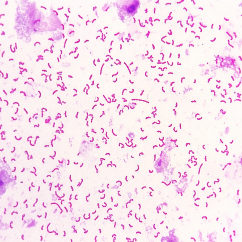
The following gram stain shows which bacteria?
Which of the following is a non-motile bacterium?
What is the most common cause of polymyositis?
Chlamydia is associated with which of the following structures?
Verotoxin-producing E. coli O157:H7 serotype belongs to which category of E. coli?
In a burn ward, multiple patients have developed Staphylococcus infections. Where is Staphylococcus typically colonized in the human body?
A 60-year-old diabetic male developed renal failure and was initiated on Chronic Ambulatory Peritoneal Dialysis (CAPD). After 4 months of CAPD, the patient developed peritonitis. What is the most common organism responsible?
Practice by Chapter
Staphylococci
Practice Questions
Streptococci and Enterococci
Practice Questions
Neisseria and Moraxella
Practice Questions
Corynebacterium and Listeria
Practice Questions
Bacillus and Clostridium
Practice Questions
Enterobacteriaceae
Practice Questions
Vibrio, Aeromonas, and Plesiomonas
Practice Questions
Pseudomonas and Related Bacteria
Practice Questions
Haemophilus and HACEK Group
Practice Questions
Bordetella and Brucella
Practice Questions
Mycobacteria
Practice Questions
Spirochetes
Practice Questions
Want unlimited practice?
Get full access to all questions, explanations, and performance tracking.
Scan to download app