
Bacteriology — MCQs

On this page
Which staphylococcal toxin is responsible for food poisoning?
Antigenic variation is/are seen in which of the following?
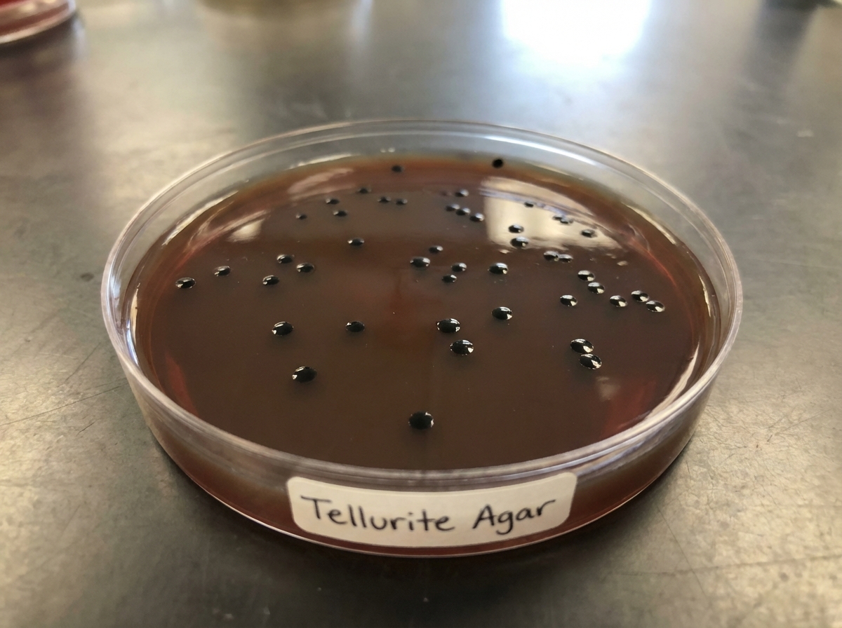
A 60-year-old male with a history of COPD and heart failure presented with shortness of breath and signs of right-sided heart failure. He was catheterized and initiated on treatment. After 8 days, he developed right flank pain, high-grade fever, and rigors. Examination revealed cloudy urine in the catheter and pain in the urethral region. Urine microscopy and culture were performed. Ultrasound of the abdomen showed pyelonephritis of the right kidney. Urine studies indicated increased WBCs. The isolated organism, when Gram stained, showed tiny deep pink colonies on MacConkey agar. Colonies grown on tellurite agar are provided. The organism is heat-resistant and can grow in 40% bile, 6.5% NaCl, and at pH 9.6. To which of the following drugs is this organism intrinsically resistant?
All of the following regarding tetanus are true except:
What is the incubation period of Salmonella in food poisoning?
All of the following are true about V. cholerae O139 except:
What is the most common organism involved in acute bacterial prostatitis?
Medusa head colonies on nutrient agar are seen in which bacterium?
Which of the following is not found in meningococci?
Which of the following is NOT true about Corynebacterium diphtheriae?
Practice by Chapter
Staphylococci
Practice Questions
Streptococci and Enterococci
Practice Questions
Neisseria and Moraxella
Practice Questions
Corynebacterium and Listeria
Practice Questions
Bacillus and Clostridium
Practice Questions
Enterobacteriaceae
Practice Questions
Vibrio, Aeromonas, and Plesiomonas
Practice Questions
Pseudomonas and Related Bacteria
Practice Questions
Haemophilus and HACEK Group
Practice Questions
Bordetella and Brucella
Practice Questions
Mycobacteria
Practice Questions
Spirochetes
Practice Questions
Want unlimited practice?
Get full access to all questions, explanations, and performance tracking.
Scan to download app