Frisch bacillus affects most commonly
Kanagawa's phenomenon is seen in ?
Subterminal spores are seen in which of the following organisms?
A militant presents with rashes all over his body sparing the palms and soles. On examination, he was febrile and lice were noted. Which of the following is responsible for his condition?
Which of the following does not possess superantigen properties?
Which organism is responsible for causing Lymphogranuloma venereum (LGV)?
Which of the following cell components produced by Neisseria gonorrhoeae is responsible for attachment to host cells?
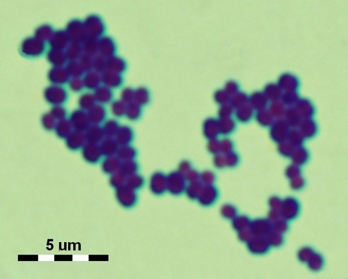
Identify the following bacterium from the image.

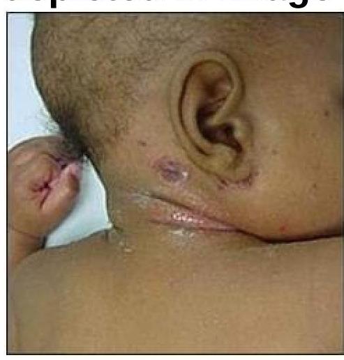
What is the causative organism for the condition depicted in the image?

A 6-year-old boy presents with fever and chills, cough, rapid breathing, difficulty breathing, and chest pain. A culture from a respiratory sample shows Gram-positive bacteria. What is the most likely organism causing this infection?
Explanation: ***Nose*** - *Klebsiella rhinoscleromatis*, also known as Frisch bacillus, is the causative agent of **rhinoscleroma**, a chronic granulomatous disease. - This bacterium primarily affects the **nasal mucosa** and upper respiratory tract, leading to progressive inflammation and nodule formation. *Mouth* - While *Klebsiella* species can be found in the oral cavity as commensals, *Klebsiella rhinoscleromatis* does not typically cause primary disease predominantly affecting the mouth. - Other infections of the mouth are more commonly linked to different bacterial or fungal pathogens. *Eye* - Ocular involvement in conditions caused by Frisch bacillus is **rare** and usually secondary to extensive spread from the primary nasal sites, not a direct primary infection. - The eye has its own specific set of infectious agents that commonly cause primary ocular pathology. *Ear* - Infection of the ear by Frisch bacillus is **not a common or primary manifestation** of rhinoscleroma. - Ear infections are typically caused by bacteria like *Streptococcus pneumoniae* or *Haemophilus influenzae*, especially in otitis media.
Explanation: ***Vibrio parahaemolyticus*** - The **Kanagawa phenomenon** refers to the production of a **thermostable direct hemolysin (TDH)** by *Vibrio parahaemolyticus*. - This hemolysin is associated with gastroenteritis caused by contaminated seafood, leading to **beta-hemolysis** on Wagatsuma agar. *Pseudomonas aeruginosa* - This bacterium is known for producing various toxins and enzymes, but the **Kanagawa phenomenon** specifically relates to hemolysin production by *Vibrio parahaemolyticus*, not *Pseudomonas aeruginosa*. - *P. aeruginosa* is often associated with **opportunistic infections** in immunocompromised patients, burn victims, and individuals with cystic fibrosis. *Shigella sonnei* - *Shigella sonnei* is a common cause of **bacterial dysentery** (shigellosis) and does not exhibit the Kanagawa phenomenon. - Its pathogenicity is primarily due to its ability to invade and replicate within colonic epithelial cells and produce **Shiga toxin**. *Proteus mirabilis* - *Proteus mirabilis* is a common cause of **urinary tract infections** and is characterized by its **swarming motility** on agar. - It does not produce the specific hemolysin associated with the Kanagawa phenomenon.
Explanation: ***Clostridium perfringens*** - This bacterium is known for producing **subterminal spores**, meaning the spores are located near, but not at, the end of the cell. - *C. perfringens* is a common cause of **gas gangrene** and food poisoning, often forming spores under adverse environmental conditions. *Clostridium tetani* - *C. tetani* produces **terminal spores**, which are found directly at one end of the bacterial cell. - These terminal spores give the organism a characteristic **"drumstick" or "tennis racket" appearance**. *Clostridium tertium* - Similar to *Clostridium tetani*, *C. tertium* is also known to produce **terminal spores**. - Its spores are typically located at the extreme end of the cell. *None of the options* - This option is incorrect because *Clostridium perfringens* specifically fits the description of producing subterminal spores. - The other *Clostridium* species listed, *C. tetani* and *C. tertium*, produce terminal spores, not subterminal.
Explanation: ***Rickettsia prowazekii*** - This bacterium is the causative agent of **epidemic typhus**, which is transmitted by the **body louse** (Pediculus humanus corporis), consistent with the presence of lice. - The characteristic rash of epidemic typhus **spares the palms and soles** and often begins on the trunk, spreading centrifugally. *Rickettsia typhi* - This organism causes **murine typhus**, which is transmitted by **fleas**, particularly those found on rodents. - While it can cause a rash that typically spares the palms and soles, the transmission vector and typical setting (exposure to rodents) do not fit the clinical scenario as well as **body lice**. *Rickettsia akari* - This bacterium causes **rickettsialpox**, transmitted by **mites**. - The rash of rickettsialpox is typically papulovesicular, and a characteristic **eschar** (or black scab) is usually present at the site of the mite bite, which is not mentioned in the case. *Rickettsia conorii* - This organism causes **Mediterranean spotted fever** (also known as Boutonneuse fever), transmitted by **ticks**. - A distinguishing feature of this infection is the presence of a **tache noire** (eschar) at the site of the tick bite, which is absent in the patient's presentation.
Explanation: ***Vibrio cholerae*** - This bacterium is primarily known for producing **cholera toxin**, which acts on the **adenylate cyclase** pathway to cause severe watery diarrhea. - While it produces powerful toxins, these do not function as **superantigens**, meaning they do not directly bind to MHC class II and T-cell receptors outside the antigen-binding groove. *Streptococcal pyrogenic exotoxins* - These toxins, produced by **Streptococcus pyogenes**, are well-known **superantigens** that cause symptoms such as **scarlet fever** and **streptococcal toxic shock syndrome**. - They bind directly to MHC class II molecules and T-cell receptors, leading to widespread T-cell activation and massive cytokine release. *Staphylococcal enterotoxins* - Produced by **Staphylococcus aureus**, these toxins are a classic example of **superantigens**, causing symptoms such as **food poisoning** and **toxic shock syndrome**. - They bypass conventional antigen presentation, nonspecifically activating a large proportion of T cells and inducing a massive inflammatory response. *None of these* - This option is incorrect because *Vibrio cholerae* stands out as the organism among the choices that does not produce superantigens. - Both streptococcal pyrogenic exotoxins and staphylococcal enterotoxins are well-established superantigens.
Explanation: ***Chlamydia trachomatis*** - Specific **serovars L1, L2, and L3** of *Chlamydia trachomatis* are responsible for causing **Lymphogranuloma venereum (LGV)**. - These serovars infect macrophages and proliferate in regional lymph nodes, leading to characteristic **buboes** and **genital ulcers**. *Neisseria gonorrhoeae* - This bacterium causes **gonorrhea**, an STI characterized by **urethritis**, cervicitis, and pharyngitis. - It does not cause the invasive lymphatic infection associated with LGV. *Treponema pallidum* - This spirochete is the causative agent of **syphilis**, which presents with **chancres**, rashes, and systemic symptoms in different stages. - Syphilis is distinct from LGV in its clinical presentation and pathology. *Haemophilus ducreyi* - This bacterium causes **chancroid**, a sexually transmitted infection resulting in painful **genital ulcers** with a ragged border and often accompanied by tender inguinal lymphadenopathy. - While it causes genital lesions and lymphadenopathy, these are distinct from the characteristic buboes and progressive lymphatic destruction of LGV.
Explanation: ***Pili (fimbriae)*** - **Pili** are hair-like appendages on the surface of *Neisseria gonorrhoeae* that facilitate initial attachment to **eukaryotic host cells**, particularly epithelial cells in the genitourinary tract. - This attachment is crucial for **colonization** and preventing the bacteria from being washed away by bodily fluids. *Lipooligosaccharide* - **Lipooligosaccharide (LOS)** is a major component of the outer membrane of *Neisseria gonorrhoeae* that contributes to **virulence** through its endotoxin activity, causing inflammation and tissue damage. - While it's important for pathogenicity, its primary role isn't direct attachment but rather the **immune response modulation** and tissue invasion. *IgA1 protease* - **IgA1 protease** is an enzyme produced by *Neisseria gonorrhoeae* that cleaves **IgA1 antibodies**, which are found on mucosal surfaces. - This enzyme helps the bacteria **evade the host immune response** by destroying protective antibodies, not directly mediating cell attachment. *Outer membrane porin protein* - **Outer membrane porin proteins (Porins)** form channels in the outer membrane of *Neisseria gonorrhoeae*, allowing the passage of small molecules. - While important for bacterial metabolism and survival, porins are not the primary structures responsible for **initial host cell attachment**.
Explanation: ⚠️ **CRITICAL IMAGE ERROR DETECTED**: The current image URL points to *Staphylococcus aureus*, but this question is about *Neisseria* species identification. The image must show Gram-negative diplococci, NOT Gram-positive cocci in clusters. This question cannot be validly answered until the correct image is provided. --- **Expected Answer (when correct image is provided):** ***Neisseria meningitidis*** - The image should display typical **Gram-negative diplococci**, which are characteristic of *Neisseria* species. - The arrangement of the bacteria in pairs strongly suggests **Neisseria meningitidis**, a common cause of bacterial meningitis. - *N. meningitidis* appears as **kidney-bean shaped diplococci** with adjacent concave sides. *Neisseria cinerea* - While also a **Gram-negative diplococcus**, *Neisseria cinerea* is generally considered non-pathogenic and rarely causes invasive disease. - Differentiation from pathogenic *Neisseria* requires **biochemical tests** or **molecular methods** beyond microscopic morphology alone. *Neisseria gonorrhoeae* - This bacterium is also a **Gram-negative diplococcus** typically associated with **gonorrhea** and other sexually transmitted infections. - Morphologically **indistinguishable** from *N. meningitidis* by light microscopy alone; definitive identification requires **culture characteristics** and **biochemical tests** (oxidase positive, glucose fermentation positive, maltose fermentation negative). *Neisseria polysaccharea* - *Neisseria polysaccharea* is a **non-pathogenic Neisseria species** found in the human upper respiratory tract. - Shares the **Gram-negative diplococci** morphology but is not a significant human pathogen and requires biochemical differentiation.
Explanation: ***Staphylococci*** - The image shows **impetigo** with **crusted lesions**, consistent with **_Staphylococcus aureus_** infection. - **Staphylococcus aureus** is a major causative organism of impetigo, particularly **bullous impetigo**, and commonly produces the characteristic **honey-colored crusts** seen in non-bullous forms as well. - This superficial bacterial skin infection is highly contagious and responds well to topical or systemic antibiotics. *Candidal infection* - **Candidal infections** (e.g., candidiasis) typically present as **erythematous patches** with satellite lesions, or white plaques in mucosal areas, which is not consistent with the image. - This fungal infection is often seen in immunocompromised individuals or in warm, moist skin folds, not as crusted superficial lesions. *Streptococcus* - While **_Streptococcus pyogenes_** can also cause impetigo (especially non-bullous impetigo), the clinical presentation in the image is most consistent with **staphylococcal infection**. - Streptococcal infections may present similarly but can also cause other conditions like cellulitis or erysipelas with distinct features. *Actinomycetes* - **Actinomycosis** is a rare, chronic bacterial infection that forms **abscesses and sinus tracts**, often with "sulfur granules," which is distinct from the superficial skin lesions shown. - This infection usually involves deeper tissues and presents as a chronic, indolent infection, unlike the acute superficial presentation of impetigo.
Explanation: ***Streptococcus pneumoniae*** - This clinical picture describes typical symptoms of **pneumonia** in a child, including fever, cough, rapid and difficult breathing, and chest pain. - **_Streptococcus pneumoniae_** is the most common bacterial cause of community-acquired pneumonia in children. The respiratory sample showing gram-positive bacteria further supports this. *Staphylococcus aureus* - While **_Staphylococcus aureus_** can cause pneumonia, it is less common than _Streptococcus pneumoniae_ in community-acquired cases in healthy children and often associated with more severe, necrotizing forms or post-viral infections. - While it is a **Gram-positive bacterium**, its clinical presentation would not be the most likely first choice for typical pneumonia symptoms in this age group. *Propionibacterium acnes* - **_Propionibacterium acnes_** (now *Cutibacterium acnes*) is primarily associated with **acne vulgaris** and, less commonly, opportunistic infections related to implanted devices or some rare soft tissue infections. - It is not a typical cause of primary respiratory infections like pneumonia. *Streptococcus pyogenes* - **_Streptococcus pyogenes_** (Group A Streptococcus) is known for causing **pharyngitis** (strep throat), skin infections (impetigo, cellulitis), and scarlet fever. - While it can rarely cause pneumonia, it is not a common cause, and the constellation of symptoms points more strongly to _Streptococcus pneumoniae_.
Staphylococci
Practice Questions
Streptococci and Enterococci
Practice Questions
Neisseria and Moraxella
Practice Questions
Corynebacterium and Listeria
Practice Questions
Bacillus and Clostridium
Practice Questions
Enterobacteriaceae
Practice Questions
Vibrio, Aeromonas, and Plesiomonas
Practice Questions
Pseudomonas and Related Bacteria
Practice Questions
Haemophilus and HACEK Group
Practice Questions
Bordetella and Brucella
Practice Questions
Mycobacteria
Practice Questions
Spirochetes
Practice Questions
Get full access to all questions, explanations, and performance tracking.
Start For Free