Which spirochete is the causative agent of syphilis?
A patient presents with sinus tracts on the foot, and a smear reveals filamentous organisms.
A 27-year-old patient presents with motheaten alopecia, moist perianal lesions, and an asymptomatic macular rash. What is the most likely causative organism?
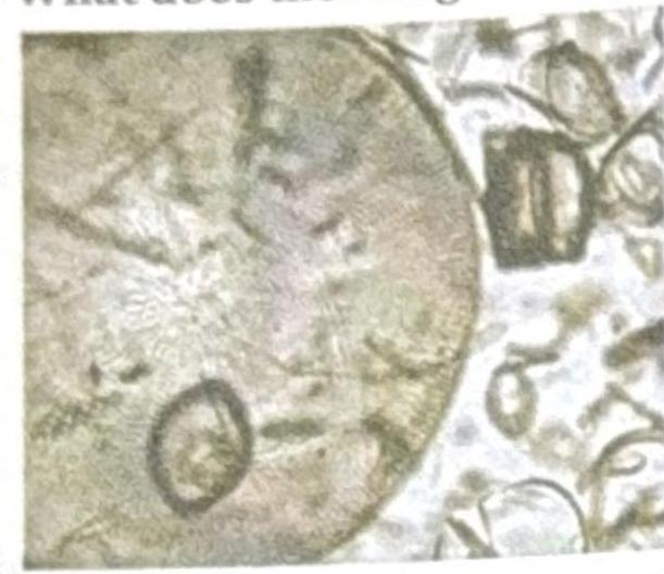
The image shows microscopic organisms. What is the most appropriate description of these organisms?

A farmer presents with a subcutaneous wound on his foot with discharge. Microscopy of a white granule from the wound shows Gram-positive filamentous rods. What is the most likely organism?
A patient was diagnosed with Escherichia coli O157:H7 infection. In this designation, what does the "H" stand for?
An adult patient with a military background is admitted with a rash, fever, altered sensorium, and a deficiency of the membrane attack complex. What is the most likely etiological agent?
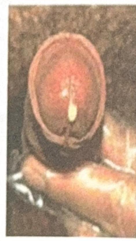
A male patient presents with white discharge from the urethra, as shown in the image. What is the most probable causative organism?

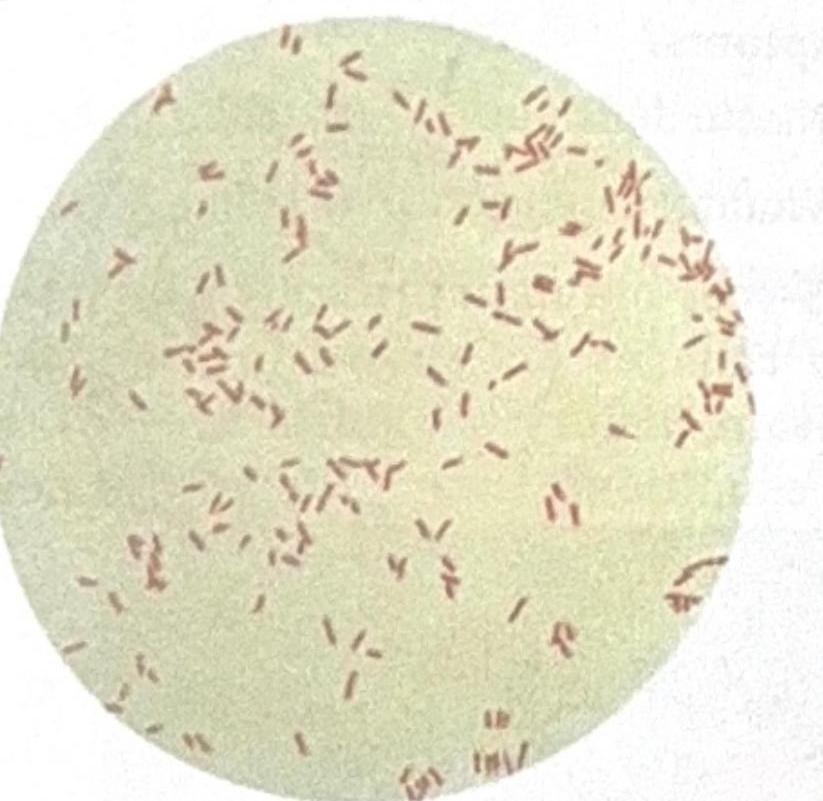
A patient presented with 70% burns, and a sample was collected from the burn site. The image shows Gram-negative rods, and the suspected organism is an obligate aerobe. What is the most likely causative microbe?

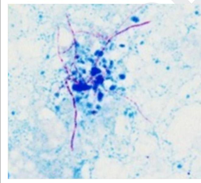
Which of the following is a gram-positive organism that shows the following appearance on Ziehl-Neelsen staining?

Explanation: ***Correct: Treponema pallidum*** - **_Treponema pallidum_** is the specific spirochete responsible for causing **syphilis**, a sexually transmitted infection. - This bacterium is characterized by its **helical shape** and corkscrew-like motility, which helps it penetrate tissues. *Incorrect: Borrelia burgdorferi* - **_Borrelia burgdorferi_** is the causative agent of **Lyme disease**, transmitted by ticks. - Its clinical presentation involves **erythema migrans**, arthritis, and neurological symptoms, distinct from syphilis. *Incorrect: Leptospira interrogans* - **_Leptospira interrogans_** causes **leptospirosis**, a zoonotic disease typically acquired through contact with contaminated water or soil. - Symptoms can range from mild flu-like illness to severe forms like **Weil's disease**, involving liver and kidney failure. *Incorrect: Borrelia recurrentis* - **_Borrelia recurrentis_** is the bacterium responsible for **louse-borne relapsing fever**. - It is transmitted by the human body louse and causes recurrent febrile episodes due to **antigenic variation**.
Explanation: ***Correct: Nocardia*** - **Nocardia species** cause **actinomycetoma**, a chronic infection characterized by **sinus tracts** discharging purulent material with sulfur granules - Smear shows **branching filamentous organisms** that are gram-positive and partially acid-fast - Classic presentation: sinus tracts on foot with filamentous organisms on direct microscopy - Key features: aerobic actinomycetes, branching at acute angles (45°) *Incorrect: Sporothrix* - Causes **sporotrichosis** (lymphocutaneous nodules along lymphatics), not sinus tracts - **Dimorphic fungus** diagnosed primarily by culture, not direct smear - Clinical presentation: nodular lesions following trauma (rose gardener's disease) - Does not show filamentous organisms on direct smear *Incorrect: Dermatophytes* - Cause **superficial infections** of skin, hair, and nails (tinea pedis, ringworm) - Do not form **deep sinus tracts** or involve subcutaneous tissue - Microscopy shows septate hyphae in skin scrapings, not in discharge from sinus tracts - Clinical presentation completely different from actinomycetoma *Incorrect: Candida* - **Yeast** causing mucocutaneous infections (thrush, vaginitis) or systemic candidiasis - Does not cause **sinus tracts** on the foot - Microscopy reveals **budding yeasts and pseudohyphae**, not true branching filaments - Not associated with actinomycetoma-type presentations
Explanation: ***Treponema*** - The constellation of **motheaten alopecia**, **moist perianal lesions** (condyloma lata), and an **asymptomatic macular rash** is highly characteristic of **secondary syphilis**, caused by *Treponema pallidum*. - **Condyloma lata** are highly infectious, raised plaques found in moist areas, and the rash can affect palms and soles. *Haemophilus* - *Haemophilus influenzae* is primarily associated with **respiratory infections** (e.g., otitis media, epiglottitis, pneumonia) and sometimes meningitis. - It does not cause the specific dermatological manifestations described in the patient. *Klebsiella* - *Klebsiella pneumoniae* is a common cause of **nosocomial infections**, particularly **pneumonia** (often with currant jelly sputum), **UTIs**, and wound infections. - It is not associated with skin conditions like alopecia or maculopapular rashes typical of syphilis. *Herpes Simplex* - Herpes Simplex Virus (HSV) causes **vesicular or ulcerative lesions**, commonly known as cold sores or genital herpes. - It does not cause motheaten alopecia, moist perianal plaques (condyloma lata), or an asymptomatic macular rash in the same manner as syphilis.
Explanation: ***Unicellular bacteria*** - The image depicts **bacteria**, which are by definition unicellular prokaryotic microorganisms - Bacteria exhibit varied morphologies (cocci, bacilli, spirilla) which can be visualized under microscopy - This is the most appropriate description among the given options for microscopic bacterial forms *Wall contains silica and chlorophyll* - Silica cell walls are characteristic of **diatoms** (photosynthetic protists), not bacteria - Bacteria have peptidoglycan cell walls, not silica - While some bacteria contain pigments, they do not contain chlorophyll in the same form as eukaryotic organisms *Present only in bone marrow* - This statement is anatomically incorrect for bacteria - Bacteria are ubiquitous and can be found in virtually all environments - Bone marrow primarily contains hematopoietic cells, not bacteria in normal conditions *Not resistant to acid* - This is not a defining characteristic that can be determined from microscopic appearance alone - Many bacteria (e.g., *Mycobacterium tuberculosis*) are **acid-fast** and highly resistant to acid - Acid resistance varies among different bacterial species and requires specific staining (Ziehl-Neelsen) to determine
Explanation: ***Nocardia*** - The presence of **white granules** in the discharge, along with **Gram-positive, filamentous rods**, is highly characteristic of *Nocardia* infection, often forming **sulfur granules** (though not always yellow). - *Nocardia* is a common soil bacterium, making it a likely pathogen in a **farmer with a subcutaneous wound** related to environmental exposure. *Staphylococcus aureus* - While *Staphylococcus aureus* can cause skin infections and abscesses, it presents as **Gram-positive cocci in clusters**, not filamentous rods. - It does not typically form **granules** in the discharge in the same manner as *Nocardia*. *Histoplasma* - *Histoplasma* is a **dimorphic fungus** that causes systemic infections, often acquired by inhaling spores. - It would appear as **yeast forms** in tissue or cultures, not Gram-positive filamentous rods, and is not typically associated with subcutaneous wounds forming granules. *Sporothrix* - *Sporothrix schenckii* causes **sporotrichosis**, characterized by a **subcutaneous nodule** that progresses along lymphatic channels. - It is a **dimorphic fungus** (yeast in tissue, mold in culture) and would not appear as Gram-positive filamentous rods on microscopy.
Explanation: **Flagella** - The "H" in bacterial designations like *Escherichia coli* O157:H7 refers to the **H antigen**, which is associated with bacterial **flagella**. - Flagellar antigens are important for serotyping bacteria, identifying different strains based on their unique flagellar protein compositions. *Lipopolysaccharide (LPS)* - The "O" in bacterial designations like *E. coli* O157 refers to the **O antigen**, which is part of the **lipopolysaccharide (LPS)** layer on the outer membrane of Gram-negative bacteria. - LPS is a major component of the bacterial outer membrane and acts as an **endotoxin**. *Capsule* - The "K" in some bacterial serotypes (e.g., *Klebsiella pneumoniae* K1) refers to the **K antigen**, which is associated with the **capsule**. - The capsule is a protective layer surrounding some bacteria that helps them evade phagocytosis. *Fimbriae* - **Fimbriae** (also known as pili) are short, hair-like appendages on the surface of some bacteria that aid in **adhesion** to host cells. - While fimbriae have their own antigenic properties, they are not typically designated by the "H" prefix in standard serotyping, which specifically refers to flagellar antigens.
Explanation: ***Neisseria meningitidis*** - A deficiency in the **membrane attack complex (MAC)**, particularly **C5-C9 components**, predisposes individuals to recurrent infections with encapsulated bacteria, especially *N. meningitidis*. - *N. meningitidis* is a common cause of **meningitis**, presenting with **fever, altered sensorium**, and often a **petechial rash** due to disseminated intravascular coagulation (DIC), which align with the patient's symptoms. *Klebsiella pneumoniae* - While *K. pneumoniae* can cause severe infections, including pneumonia and meningitis, it is not specifically associated with **MAC deficiency**. - Its infections more commonly manifest as **severe pneumonia** or **urinary tract infections** in immunocompromised patients. *Haemophilus influenzae* - *H. influenzae* can cause meningitis and other invasive infections, especially in children, but it is not typically linked to **MAC deficiency**. - The classic presentation involving **rash** and severe systemic symptoms as described is more characteristic of **meningococcal disease**. *CMV* - **Cytomegalovirus (CMV)** is a herpesvirus that causes a wide range of diseases, particularly in immunocompromised individuals. - However, CMV infections are primarily associated with **cellular immunity defects** rather than a deficiency in the **membrane attack complex** of the complement system.
Explanation: ***Neisseria gonorrhoeae*** - Among the given options, *Neisseria gonorrhoeae* is the **most probable causative organism** for **urethral discharge** in males. - Gonorrhea is a common sexually transmitted infection (STI) presenting with **purulent urethritis**, typically with thick, yellow-green discharge, though appearance can vary. - **Clinical note:** While classic gonococcal discharge is purulent and yellow-green, the clinical presentation can vary. None of the other organisms listed cause urethritis with discharge. *Haemophilus ducreyi* - This bacterium causes **chancroid**, a sexually transmitted infection characterized by **painful genital ulcers (chancres)** and **inguinal lymphadenopathy**, not urethral discharge. - Presents with ulcerative lesions, not discharge. *Klebsiella granulomatis* - This organism is responsible for **donovanosis** (granuloma inguinale), which manifests as **painless, progressive ulcerative lesions** on the genitals. - It does not cause urethral discharge; presents with beefy red granulomatous lesions. *Treponema pallidum* - This spirochete causes **syphilis**, which presents with **painless chancres** in the primary stage, **maculopapular rash** in the secondary stage, and gummas or neurological symptoms in later stages. - Urethral discharge is not a typical symptom of syphilis; primary lesions are ulcerative.
Explanation: ***Pseudomonas aeruginosa*** - The image shows **Gram-negative rods**, and the patient has extensive **burns**, making *Pseudomonas aeruginosa* a highly likely causative agent due to its common association with burn wound infections. - *Pseudomonas aeruginosa* is an **obligate aerobe** and thrives in moist environments, making it a frequent colonizer of burn wounds, which are large, often moist surfaces. *Neisseria meningitidis (Meningococcus)* - *Neisseria meningitidis* is a **Gram-negative coccus**, typically appearing as diplococci, not rods, on Gram stain. - While it can cause severe infections, it is primarily associated with **meningitis** and **sepsis**, not typically burn wound infections. *Streptococcus pneumoniae (Pneumococcus)* - *Streptococcus pneumoniae* is a **Gram-positive coccus**, appearing as lancet-shaped diplococci or short chains, which contradicts the Gram-negative rod morphology seen in the image. - It is a common cause of **pneumonia** and **otitis media**, not primarily associated with burn wound infections. *Streptococcus pyogenes* - *Streptococcus pyogenes* is a **Gram-positive coccus** that grows in chains, which is inconsistent with the Gram-negative rod morphology. - Although it can cause skin infections like cellulitis and impetigo, it is not a typical cause of **burn wound infections** in the way *Pseudomonas aeruginosa* is.
Explanation: ***Nocardia*** * The image displays delicate, branching, **filamentous rods** that are stained **red/pink** against a blue background, which is characteristic of partially acid-fast organisms like *Nocardia* on a Ziehl-Neelsen stain. * *Nocardia* species are **gram-positive**, aerobic bacteria that can cause opportunistic infections, particularly in immunocompromised individuals. They are distinguished by their **partial acid-fastness** due to their mycolic acid content, similar to mycobacteria but to a lesser degree. * The characteristic **branching filamentous morphology** combined with partial acid-fastness on Ziehl-Neelsen staining is pathognomonic for *Nocardia*. *Incorrect: Mycobacterium tuberculosis* * While *M. tuberculosis* is **strongly acid-fast** on Ziehl-Neelsen staining (appearing red), it is **not truly gram-positive**—it is gram-variable or weakly gram-positive. * *Mycobacterium* appears as **straight or slightly curved rods**, NOT branching filaments like those shown in the image. *Incorrect: Actinomyces* * *Actinomyces* is a **gram-positive**, filamentous, branching organism that can morphologically resemble *Nocardia*. * However, *Actinomyces* is **NOT acid-fast** and would appear **blue** (not red/pink) on Ziehl-Neelsen staining as it takes up the counterstain. * *Actinomyces* is also anaerobic, whereas *Nocardia* is aerobic. *Incorrect: Rhodococcus* * *Rhodococcus* is a gram-positive organism that can show **partial acid-fastness**, similar to *Nocardia*. * However, *Rhodococcus* typically appears as **coccoid to short rods**, occasionally forming short chains, but does NOT show the extensive **branching filamentous** pattern characteristic of *Nocardia* seen in the image.
Staphylococci
Practice Questions
Streptococci and Enterococci
Practice Questions
Neisseria and Moraxella
Practice Questions
Corynebacterium and Listeria
Practice Questions
Bacillus and Clostridium
Practice Questions
Enterobacteriaceae
Practice Questions
Vibrio, Aeromonas, and Plesiomonas
Practice Questions
Pseudomonas and Related Bacteria
Practice Questions
Haemophilus and HACEK Group
Practice Questions
Bordetella and Brucella
Practice Questions
Mycobacteria
Practice Questions
Spirochetes
Practice Questions
Get full access to all questions, explanations, and performance tracking.
Start For Free