
Bacteriology — MCQs

On this page
Fermentation of glycerol is the basis for classification of:
Gas gangrene can be caused by all Except
Which of the following is not a biovar of Corynebacterium diphtheriae?
Bacteria most commonly involved in prosthetic valvular heart disease within 2 months of surgery is:
The term 'leathery' appearance in medical terminology is characteristically used to describe:
In a patient of the nephrotic syndrome with spontaneous bacterial peritonitis, which one of the following micro-organisms is most commonly involved?
All of the following statements about cholera are true except -
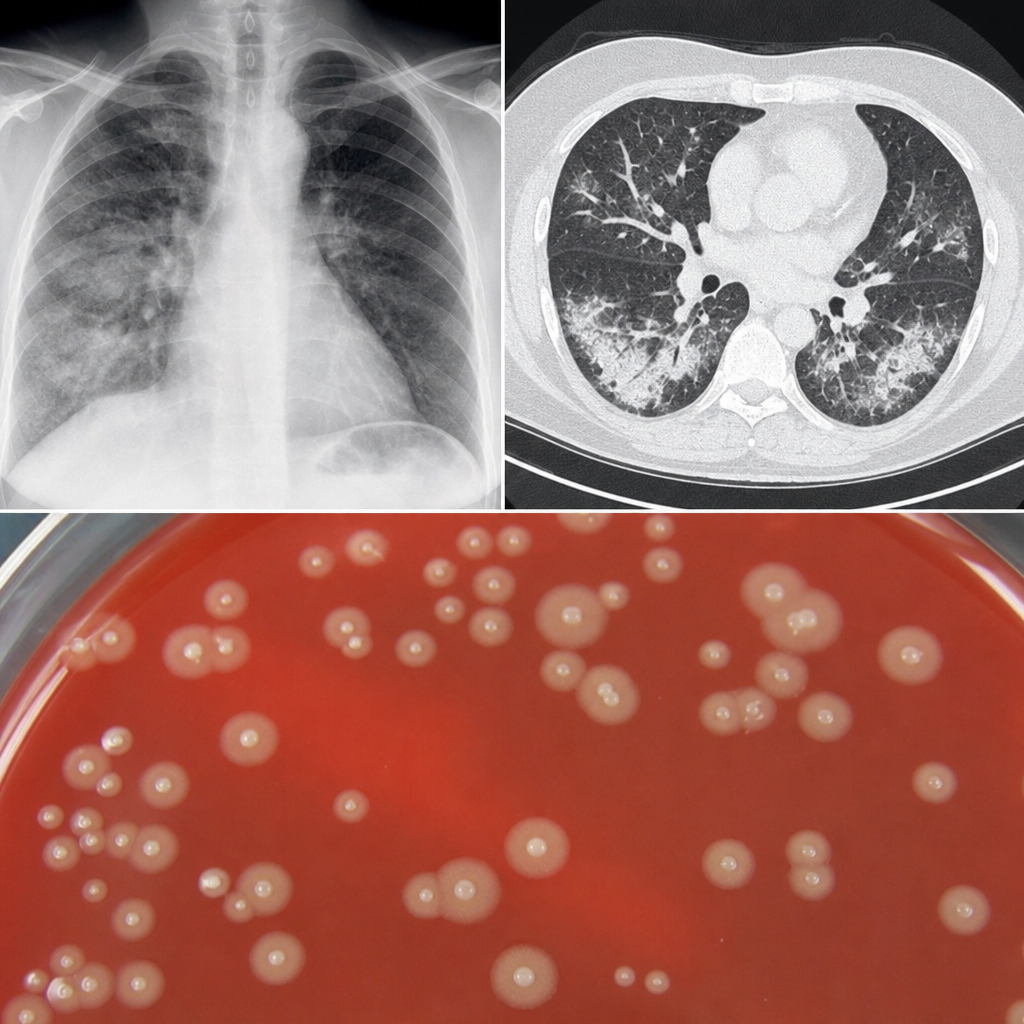
A 40-year-old woman complained of unrelenting headache, accompanied by fever, chills, and malaise. After 3 to 4 days, a dry cough developed. Her white cell count was normal. Chest x-ray and CT scan were done. A pathogen I suspected which produces colonies like the ones given below. Which of the following is the most likely diagnosis?
“Hundred day cough” is the name of
A 4-year-old male child presents with fever, anemia and azotemia after an episode of dysentery 9 days earlier. The commonest organism responsible for this condition is?
Practice by Chapter
Staphylococci
Practice Questions
Streptococci and Enterococci
Practice Questions
Neisseria and Moraxella
Practice Questions
Corynebacterium and Listeria
Practice Questions
Bacillus and Clostridium
Practice Questions
Enterobacteriaceae
Practice Questions
Vibrio, Aeromonas, and Plesiomonas
Practice Questions
Pseudomonas and Related Bacteria
Practice Questions
Haemophilus and HACEK Group
Practice Questions
Bordetella and Brucella
Practice Questions
Mycobacteria
Practice Questions
Spirochetes
Practice Questions
Want unlimited practice?
Get full access to all questions, explanations, and performance tracking.
Scan to download app