
Bacteriology — MCQs

On this page
HP bodies (Halberstaedter-Prowazek bodies) are seen in infections caused by which organism?
A 20-year-old man presents with dysuria, urgency, and urethral discharge. Physical examination shows suppurative urethritis, with redness and swelling at the urethral meatus. Which of the following is the most likely etiology of urethritis in this patient?
Which organism causes prosthetic valve endocarditis within 60 days of surgery?
In which year was Helicobacter pylori identified?
Mouse is used for pathogenicity testing in -
True about Trench fever are all except -
Vibrio parahemolyticus food poisoning is caused by ingestion of -
Chancroid may be caused by:
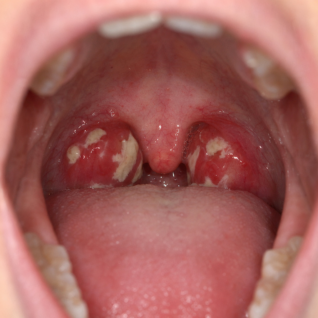
Most common bacteria responsible for the condition shown in the image below in a child who presents with high grade fever and sore throat is?
Most common agent causing tuberculosis in AIDS patients in tropical countries is:
Practice by Chapter
Staphylococci
Practice Questions
Streptococci and Enterococci
Practice Questions
Neisseria and Moraxella
Practice Questions
Corynebacterium and Listeria
Practice Questions
Bacillus and Clostridium
Practice Questions
Enterobacteriaceae
Practice Questions
Vibrio, Aeromonas, and Plesiomonas
Practice Questions
Pseudomonas and Related Bacteria
Practice Questions
Haemophilus and HACEK Group
Practice Questions
Bordetella and Brucella
Practice Questions
Mycobacteria
Practice Questions
Spirochetes
Practice Questions
Want unlimited practice?
Get full access to all questions, explanations, and performance tracking.
Scan to download app