A female child presents with virilization and hypertension with low plasma renin. What is the most likely diagnosis?
Which of the following statements is false regarding Menkes disease?
What is the most common cardiac defect in Turner syndrome?
Locus heterogeneity is a feature of which of the following?
All of the following diseases are associated with trinucleotide repeat sequences except?
What is the term for a single gene defect that causes multiple, seemingly unrelated problems?
Schwannoma of spinal nerve roots is typically seen in which condition?
What is the most sensitive investigation for cystic fibrosis?
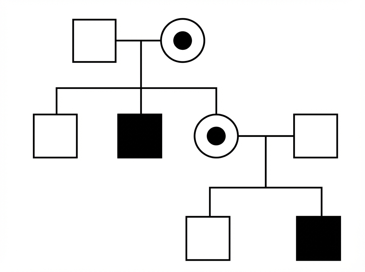
Study the following pedigree. What is the inheritance pattern of the disease in the family?

DNA topoisomerase 1 autoantibody is specific for which condition?
Explanation: The clinical presentation of **virilization** (ambiguous genitalia/androgen excess) combined with **hypertension** and **low renin** is the classic hallmark of **11β-hydroxylase deficiency**. [1] **1. Why 11β-hydroxylase deficiency is correct:** In this condition, the enzyme 11β-hydroxylase is deficient, blocking the conversion of 11-deoxycortisol to cortisol and 11-deoxycorticosterone (DOC) to corticosterone. This leads to: * **Androgen Excess:** Shunting of precursors into the androgen pathway causes virilization in females. [1] * **Hypertension:** The block leads to an accumulation of **11-deoxycorticosterone (DOC)**. DOC is a potent mineralocorticoid; its excess causes sodium retention and volume expansion, resulting in hypertension and feedback suppression of **Renin**. [1] **2. Why other options are incorrect:** * **21α-hydroxylase deficiency:** This is the most common cause of CAH. While it causes virilization, it leads to **hypotension** and "salt-wasting" because mineralocorticoids (aldosterone) cannot be produced. * **3β-hydroxysteroid dehydrogenase deficiency:** This rare form results in a lack of all adrenal steroids (glucocorticoids, mineralocorticoids, and sex steroids). It typically presents with salt-wasting and ambiguous genitalia in males (incomplete masculinization). * **Conn’s Syndrome:** While it presents with hypertension and low renin (primary hyperaldosteronism), it **does not cause virilization** as it involves only the zona glomerulosa. **High-Yield Clinical Pearls for NEET-PG:** * **11β-hydroxylase deficiency:** Hypertension + Virilization (The "1" in 11 stands for "Hypertension"). [1] * **21α-hydroxylase deficiency:** Hypotension + Virilization. [1] * **17α-hydroxylase deficiency:** Hypertension + No Virilization (Delayed puberty/Sexual infantilism). * **Key Marker:** Elevated **11-deoxycortisol** and **11-deoxycorticosterone** are diagnostic for 11β-hydroxylase deficiency.
Explanation: **Explanation:** The correct answer is **C** because Menkes disease is caused by a mutation in the **ATP7A gene**, not ATP7B. The ATP7A protein is responsible for the intestinal absorption of copper and its transport across the blood-brain barrier. In Menkes disease, copper is trapped within intestinal cells and cannot reach the bloodstream, leading to a severe **systemic copper deficiency**. In contrast, **ATP7B** mutations cause **Wilson disease**, where copper cannot be excreted into bile, leading to copper toxicity and overload. **Analysis of other options:** * **Option A (X-linked recessive):** This is a true statement. Menkes disease primarily affects males, while females are typically asymptomatic carriers. * **Option B (Mental retardation):** This is true. Low copper levels impair the function of copper-dependent enzymes like *Cytochrome c oxidase*, leading to defective energy metabolism in the brain, resulting in neurodegeneration and developmental delay. * **Option C (Kinky hair):** This is a hallmark clinical feature (Pili torti). It occurs due to a deficiency in *Lysyl oxidase*, a copper-dependent enzyme required for cross-linking keratin and collagen [1]. **NEET-PG High-Yield Pearls:** * **Enzyme Deficiency:** Low *Lysyl oxidase* leads to kinky hair and connective tissue laxity [1]; low *Tyrosinase* leads to hypopigmentation. * **Diagnosis:** Low serum copper and low serum ceruloplasmin (similar to Wilson’s, but clinical presentation is opposite). * **Treatment:** Early administration of copper histidine (parenteral) may help, but the prognosis is generally poor.
Explanation: **Explanation:** **Turner Syndrome (45, XO)** is characterized by a high prevalence of congenital heart diseases (CHD), occurring in approximately 25–50% of patients. These defects primarily involve the **left-sided heart structures**. 1. **Why Coarctation of the Aorta is Correct:** **Coarctation of the aorta** is the most common *clinically significant* cardiac defect in Turner syndrome, occurring in about 15–20% of cases. It typically presents as a narrowing of the aortic arch distal to the origin of the left subclavian artery [1]. However, it is important to note that **Bicuspid Aortic Valve (BAV)** is actually the most common overall cardiac anomaly (up to 30%), but among the options provided, Coarctation is the classic and correct answer. 2. **Why Incorrect Options are Wrong:** * **Ventricular Septal Defect (VSD) & Atrial Septal Defect (ASD):** These are common in the general population and other trisomies (like Down Syndrome/Trisomy 21), but they are not the hallmark of Turner syndrome. * **Tetralogy of Fallot (ToF):** This is a cyanotic heart disease most commonly associated with **DiGeorge Syndrome** (22q11 deletion), not Turner syndrome. **Clinical Pearls for NEET-PG:** * **The "Left-Sided" Rule:** Always associate Turner syndrome with left-sided obstructive lesions (BAV, Coarctation, Aortic Stenosis). * **Physical Exam:** Look for "radio-femoral delay" and a BP discrepancy between upper and lower limbs in a female with short stature and webbed neck [1]. * **Aortic Dissection:** Patients with Turner syndrome have a significantly increased risk of aortic root dilation and dissection, even in the absence of hypertension. * **Karyotype:** 45, XO is the most common, but mosaicism (e.g., 45,X/46,XX) can also occur.
Explanation: Locus heterogeneity occurs when mutations at different genetic loci (different genes) produce the same clinical phenotype. In the context of Muscular Dystrophies, this is a classic feature [2]. For example, while Duchenne and Becker muscular dystrophies are X-linked (mutations in the DMD gene) [1], various Limb-Girdle Muscular Dystrophies (LGMD) can be inherited in either autosomal dominant or recessive patterns due to mutations in entirely different genes (e.g., CAPN3, DYSF, or SGCG), yet they present with similar clinical muscle wasting. Analysis of Options: Muscular Dystrophies (Correct): As noted, multiple distinct gene loci can lead to the clinical syndrome of muscular dystrophy, making it a prime example of locus heterogeneity [2]. Familial Adenomatous Polyposis (Incorrect): This is primarily associated with mutations in a single gene, the APC gene on chromosome 5q21. When a single gene mutation results in different clinical manifestations, it is called allelic heterogeneity, not locus heterogeneity. Osteopetrosis (Incorrect): While there are different types (infantile vs. adult), it is generally not used as the classic textbook example for locus heterogeneity in the same way muscular dystrophies or Albinism/Retinitis Pigmentosa are. NEET-PG High-Yield Pearls: 1. Locus Heterogeneity: Different genes → Same disease (e.g., Osteogenesis Imperfecta, Albinism, Sensorineural hearing loss). 2. Allelic Heterogeneity: Different mutations in the same gene → Same disease (e.g., Beta-thalassemia, Cystic Fibrosis). 3. Pleiotropy: One gene mutation → Multiple, seemingly unrelated phenotypic effects (e.g., Marfan Syndrome affecting heart, eyes, and skeleton). 4. Variable Expressivity: Same genotype → Different degrees of severity in phenotype.
Explanation: **Explanation:** The correct answer is **Hereditary motor and sensory neuropathy (HMSN)**, also commonly known as **Charcot-Marie-Tooth (CMT) disease**. Unlike the other options, HMSN is not a trinucleotide repeat disorder. It is most commonly caused by a **duplication** of the PMP22 gene (CMT1A) on chromosome 17, or by point mutations affecting peripheral myelin proteins. **Trinucleotide Repeat Disorders** are characterized by the expansion of specific three-nucleotide sequences. When these repeats exceed a certain threshold, they cause gene instability and disease. * **Huntington’s Disease (Option A):** An autosomal dominant neurodegenerative disorder caused by **CAG** repeats (Polyglutamine disease) on Chromosome 4 [1]. * **Friedreich’s Ataxia (Option B):** An autosomal recessive spinocerebellar degeneration caused by **GAA** repeats in the Frataxin gene on Chromosome 9 [1]. * **Myotonic Dystrophy (Option C):** An autosomal dominant muscular dystrophy (Type 1) caused by **CTG** repeats in the DMPK gene on Chromosome 19. **High-Yield Clinical Pearls for NEET-PG:** 1. **Anticipation:** This is a hallmark of trinucleotide repeat diseases where the disease becomes more severe or has an earlier onset in successive generations due to further expansion of the repeats. 2. **Location of Repeats:** * **Exonic (Coding):** Huntington’s (CAG). * **Intronic (Non-coding):** Friedreich’s Ataxia (GAA). * **Untranslated Region (3' UTR):** Myotonic Dystrophy (CTG). * **Untranslated Region (5' UTR):** Fragile X Syndrome (CGG). 3. **Mnemonic for Friedreich's Ataxia:** **GAA** = **G**ait **A**taxia **A**lways.
Explanation: **Explanation:** **1. Why Pleiotropism is Correct:** **Pleiotropism** occurs when a single gene mutation results in multiple, diverse phenotypic effects across different organ systems [1]. These effects often seem unrelated but share a common underlying biochemical or structural defect. * **Classic Example:** **Marfan Syndrome**. A mutation in the *FBN1* gene (encoding Fibrillin-1) leads to defects in the skeletal system (arachnodactyly), the ocular system (ectopia lentis), and the cardiovascular system (aortic aneurysm). **2. Why Other Options are Incorrect:** * **Pseudodominance:** This occurs when a recessive trait mimics a dominant pattern of inheritance. This happens when a person homozygous for a recessive trait mates with a heterozygote carrier, resulting in a 50% chance of affected offspring in every generation. * **Penetrance:** This refers to the percentage of individuals with a specific genotype who actually express the associated phenotype. If 100 people have the gene but only 80 show symptoms, it is "incomplete penetrance." * **Anticipation:** This is the phenomenon where a genetic disorder becomes more severe or has an earlier age of onset in successive generations. It is typically seen in **Trinucleotide Repeat Disorders** (e.g., Huntington’s disease, Fragile X syndrome). **3. Clinical Pearls for NEET-PG:** * **Variable Expressivity:** Unlike penetrance (all-or-none), this refers to the *degree* or severity of the phenotype among individuals with the same genotype. * **Locus Heterogeneity:** Mutations at different loci (different genes) produce the same phenotype (e.g., Albinism, Osteogenesis Imperfecta). * **Allelic Heterogeneity:** Different mutations within the *same* gene produce the same phenotype (e.g., Cystic Fibrosis, Beta-thalassemia). * **High-Yield Pleiotropy Example:** **Phenylketonuria (PKU)** causes mental retardation, reduced hair pigmentation, and a "mousy" odor.
Explanation: The correct answer is **Neurofibromatosis type 1 (NF1)**. While NF1 is classically associated with neurofibromas (plexiform and cutaneous), it is also the most common genetic predisposition for **spinal nerve root schwannomas**. In NF1, these tumors often involve the dorsal roots and can lead to spinal cord compression. **Analysis of Options:** * **Neurofibromatosis type 1 (NF1):** Caused by a mutation in the *NF1* gene on chromosome 17 (Neurofibromin). It is characterized by Café-au-lait spots, Lisch nodules, and various nerve sheath tumors, including spinal schwannomas. * **Neurofibromatosis type 2 (NF2):** While NF2 is famous for **bilateral vestibular schwannomas** (Acoustic neuromas), spinal schwannomas are actually less frequent in NF2 compared to NF1 in the context of general spinal root involvement [1]. NF2 is more specifically associated with "MISME" (Multiple Inherited Schwannomas, Meningiomas, and Ependymomas). * **Turcot Syndrome:** A variant of FAP or HNPCC associated with CNS tumors, most commonly medulloblastoma or glioblastoma multiforme, not schwannomas. * **Li-Fraumeni Syndrome:** Caused by a *p53* mutation; it predisposes to SBLA syndrome (Sarcoma, Breast, Leukemia, Adrenal cortical carcinoma) and brain tumors, but not typically schwannomas. **High-Yield Clinical Pearls for NEET-PG:** * **NF1 (Chromosome 17):** 17 letters in "Neurofibromatosis." Look for Optic gliomas and Pheochromocytoma. * **NF2 (Chromosome 22):** 22 for "Acoustic" (2 ears/8th nerve). Look for bilateral cataracts (posterior subcapsular). * **Schwannoma Pathology:** Characterized by **Antoni A** (dense cells, Verocay bodies) and **Antoni B** (loose stroma) patterns. They are S100 positive.
Explanation: **Explanation:** **Cystic Fibrosis (CF)** is an autosomal recessive disorder caused by mutations in the **CFTR gene** (Cystic Fibrosis Transmembrane Conductance Regulator). This defect leads to impaired chloride transport across epithelial cells, resulting in thick, viscid secretions in the lungs and pancreas, and abnormally high salt content in sweat [1]. **Why Sweat Electrolytes is the Correct Answer:** The **Pilocarpine Iontophoresis Sweat Test** remains the **gold standard and most sensitive investigation** for diagnosing CF. It measures the concentration of chloride in sweat. A chloride level **>60 mmol/L** on two separate occasions is diagnostic. It is preferred because the physiological defect in CFTR is most consistently expressed in the sweat glands, where chloride cannot be reabsorbed from the primary sweat. **Why Other Options are Incorrect:** * **A & D (CT Scan/Tomogram):** While high-resolution CT (HRCT) is excellent for detecting bronchiectasis (a common complication of CF), it is not a diagnostic tool for the underlying genetic defect [1]. It shows the damage, not the disease itself. * **B (Ultrasound):** Ultrasound may show fatty infiltration of the pancreas or "meconium ileus" in neonates, but it lacks the sensitivity and specificity required for a definitive diagnosis of CF. **High-Yield Clinical Pearls for NEET-PG:** * **Screening:** Immunoreactive Trypsinogen (IRT) is used for newborn screening. * **Most Common Mutation:** ΔF508 (a class II defect involving protein misfolding). * **Nasal Potential Difference:** Used as a diagnostic tool if sweat tests are borderline. * **Common Pathogens:** *Staphylococcus aureus* (early childhood) and *Pseudomonas aeruginosa* (most common in adults). * **Infertility:** 95% of males are infertile due to Congenital Bilateral Absence of the Vas Deferens (CBAVD) [1].
Explanation: ***X-linked recessive type*** - **Affected males** appear across multiple generations connected through **unaffected carrier females**, which is characteristic of X-linked recessive inheritance. - There is **no male-to-male transmission** since fathers cannot pass their X chromosome to sons, and **daughters of affected males** are obligate carriers. *Autosomal recessive type* - Would require **both parents to be carriers** for affected offspring, typically showing affected individuals in a single generation (horizontal pattern). - **Consanguinity** is often present, and both **males and females** are equally affected, which doesn't match the pedigree pattern. *Autosomal dominant type* - Would show **vertical transmission** with affected individuals in every generation having at least one affected parent. - **Male-to-male transmission** would be possible, and approximately **50% of offspring** from affected parents would be affected. *X-Linked dominant type* - **All daughters** of affected males would be affected since they must inherit the father's X chromosome. - Affected females would have **more severely affected offspring** than the pattern typically shows, and there would be more affected females overall.
Explanation: The correct answer is **Diffuse scleroderma**. **Understanding the Concept:** DNA topoisomerase 1 autoantibody, historically known as **Anti-Scl-70**, is a highly specific marker for the **diffuse cutaneous systemic sclerosis (dcSSc)** subtype of scleroderma. Topoisomerase 1 is a nuclear enzyme responsible for relieving torsional strain during DNA replication [1]. In patients with diffuse scleroderma [2], antibodies against this enzyme are associated with extensive skin involvement and a higher risk of **interstitial lung disease (ILD)**. **Analysis of Options:** * **Limited cutaneous systemic sclerosis:** This condition is more characteristically associated with **Anti-centromere antibodies**. It typically presents with the CREST syndrome (Calcinosis, Raynaud’s, Esophageal dysmotility, Sclerodactyly, Telangiectasia) and carries a higher risk of pulmonary arterial hypertension (PAH) rather than ILD. * **Mixed connective tissue disease (MCTD):** The hallmark serological marker for MCTD is high titers of **Anti-U1 RNP (ribonucleoprotein)** antibodies [1]. * **Systemic lupus erythematosus (SLE):** While SLE involves various antibodies, the most specific are **Anti-dsDNA** [3] and **Anti-Smith (Sm)** antibodies [1]. **NEET-PG High-Yield Pearls:** * **Anti-Scl-70 (Topoisomerase 1):** Specific for Diffuse Scleroderma; predicts severe lung fibrosis. * **Anti-Centromere:** Specific for Limited Scleroderma (CREST); predicts pulmonary hypertension. * **Anti-RNA Polymerase III:** Associated with diffuse skin involvement and a high risk for **Scleroderma Renal Crisis**. * **Anti-Jo-1:** Most common antibody in Polymyositis/Dermatomyositis (specifically Antisynthetase syndrome).
Principles of Medical Genetics
Practice Questions
Genetic Testing and Counseling
Practice Questions
Single Gene Disorders
Practice Questions
Chromosomal Disorders
Practice Questions
Mitochondrial Diseases
Practice Questions
Pharmacogenomics
Practice Questions
Cancer Genetics
Practice Questions
Genetics of Common Diseases
Practice Questions
Epigenetics and Disease
Practice Questions
Genetic Basis of Developmental Disorders
Practice Questions
Ethical Issues in Medical Genetics
Practice Questions
Gene Therapy and Precision Medicine
Practice Questions
Get full access to all questions, explanations, and performance tracking.
Start For Free