Which of the following conditions is inherited in an autosomal dominant pattern?
Klinefelter syndrome is diagnosed by?
An albino girl marries a phenotypically normal boy. What are the chances of their having an affected child, and what are the chances of their children being carriers?
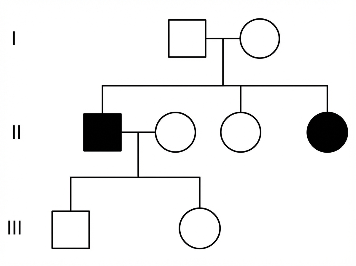
A pedigree chart shows the following pattern of inheritance. What is the most likely mode of inheritance?

Which of the following is an X-linked dominant condition?
Which enzyme levels are increased in Duchenne's muscular dystrophy?
A 32-year-old woman presents with a 2-day history of decreased vision and photopsia in her left eye. She is noted to be unusually tall with long limbs and slender fingers. Physical examination reveals pectus excavatum, kyphoscoliosis, a midsystolic click, and a pansystolic murmur. Ophthalmologic examination shows a retinal detachment and lens dislocation. The patient is at greatest risk for which of the following conditions?
All are manifestations of tuberous sclerosis, EXCEPT:
What is the commonest mode of inheritance of von Willebrand's disease?
A very tall, slender 16-year-old boy is referred for evaluation of an abnormal CXR. He reports no pulmonary or cardiac symptoms and feels well. On physical examination, he has long fingers, pectus excavatum, and a high arched palate. Which of the following is most likely to be seen on his CXR?
Explanation: **Explanation:** **Neurofibromatosis (NF)** is the correct answer as both NF Type 1 (von Recklinghausen disease) and NF Type 2 are classic examples of **Autosomal Dominant (AD)** inheritance. NF1 is caused by a mutation in the *NF1* gene on chromosome 17, while NF2 involves the *NF2* gene on chromosome 22. A key feature of AD inheritance seen in NF is **high penetrance but variable expressivity**, meaning almost everyone with the mutation shows signs, but the severity varies significantly. **Analysis of Incorrect Options:** * **G6PD Deficiency:** This is an **X-linked recessive** disorder. It primarily affects males, causing episodic hemolysis triggered by oxidative stress (e.g., fava beans, primaquine). * **Hirschsprung Disease:** This condition exhibits **complex/multifactorial inheritance**. While associated with mutations in the *RET* proto-oncogene, it does not follow a simple Mendelian AD pattern and often shows variable penetrance. * **Vitamin D Resistant Rickets (Hereditary Hypophosphatemic Rickets):** This is the classic example of **X-linked Dominant** inheritance. It is characterized by phosphate wasting in the kidneys. **High-Yield Clinical Pearls for NEET-PG:** * **NF1 Mnemonic:** "17 letters in von Recklinghausen" = Chromosome **17**. Look for Lisch nodules, Café-au-lait spots, and optic gliomas. * **NF2 Mnemonic:** "22" = Chromosome **22**. Look for bilateral acoustic neuromas (vestibular schwannomas) [1]. * **AD Inheritance Rule:** Usually involves mutations in **structural proteins** (e.g., Collagen, Fibrillin) or **receptors**, whereas Autosomal Recessive conditions usually involve **enzyme deficiencies**.
Explanation: **Explanation:** **1. Why Karyotyping is the Correct Answer:** Klinefelter syndrome is a chromosomal numerical abnormality, most commonly characterized by a **47, XXY** genotype. Since the definitive diagnosis depends on identifying the presence of an extra X chromosome in a male, **Karyotyping** (chromosomal analysis) is the gold standard. It allows for the visualization of the entire set of chromosomes to confirm the aneuploidy and rule out mosaicism (e.g., 46,XY/47,XXY). **2. Why Other Options are Incorrect:** * **USG Abdomen:** While it may show small, atrophic testes or cryptorchidism, it cannot provide a genetic diagnosis. * **Triple Test:** This is a maternal screening tool used during pregnancy to assess the risk of Trisomy 21, 18, and neural tube defects; it is not a diagnostic tool for Klinefelter syndrome. * **Echocardiography:** Although Klinefelter patients have a slightly higher risk of Mitral Valve Prolapse (MVP), this is a supportive finding, not a diagnostic one. **3. High-Yield Clinical Pearls for NEET-PG:** * **Clinical Features:** Tall stature, eunuchoid body habitus, gynecomastia, small firm testes (testicular dysgenesis), and infertility (azoospermia). * **Hormonal Profile:** **High FSH and LH** (due to loss of feedback inhibition) and **Low Testosterone** (Hypergonadotropic Hypogonadism). * **Histology:** Characterized by **Hyalinization and fibrosis of seminiferous tubules** and Leydig cell hyperplasia. * **Barr Body:** Positive (unlike normal males). * **Associated Risks:** Increased risk of Male Breast Cancer, Germ cell tumors (mediastinal), and Autoimmune diseases (SLE).
Explanation: ### Explanation **1. Understanding the Inheritance Pattern** Albinism (specifically Oculocutaneous Albinism) is a classic **Autosomal Recessive (AR)** disorder. For an individual to be affected, they must possess two mutant alleles (genotype: **aa**). A phenotypically normal individual, unless specified as a carrier, is assumed to be homozygous dominant (genotype: **AA**) in standard genetic problems. Genetic risk for carriers is often calculated based on pedigree and prior probability [1]. * **Mother (Albino):** Genotype **aa** * **Father (Normal):** Genotype **AA** Using a Punnett Square, all offspring will receive one 'a' from the mother and one 'A' from the father, resulting in a **100% Aa (Heterozygous)** genotype. Phenotypically, they are all normal, but genetically, they are all carriers. **2. Analysis of Incorrect Options** * **Option B (All normal):** While phenotypically true, it is incomplete as it ignores the carrier status, which is a critical genetic distinction. * **Option C (50% carriers):** This would only occur if the mother was a carrier (Aa) and the father was normal (AA). * **Option D (50% affected, 50% carriers):** This occurs in a **Test Cross** scenario where an affected individual (aa) mates with a carrier (Aa). Since the father is "phenotypically normal" and not specified as a carrier, we assume the general population genotype (AA). **3. NEET-PG Clinical Pearls** * **Oculocutaneous Albinism (OCA):** Most commonly due to a deficiency in the enzyme **Tyrosinase**, which converts Tyrosine to DOPA and Melanin. Diagnosis of such Inborn Errors of Metabolism (IEM) can be confirmed through biochemical genetics laboratories [1]. * **AR Inheritance Rule:** If both parents are carriers (Aa x Aa), the risk is: 25% affected, 50% carriers, and 25% normal. * **Consanguinity:** Always suspect AR disorders in questions mentioning "marriage between cousins." * **Associated Syndromes:** Look for **Chediak-Higashi Syndrome** (albinism + giant granules in neutrophils + recurrent infections).
Explanation: ***Autosomal recessive*** - Characterized by **unaffected carrier parents** producing affected offspring, with the trait **skipping generations**. - Affects **both sexes equally** and typically shows a **horizontal pattern** within a single generation. *Autosomal dominant* - Shows a **vertical pattern** where affected individuals have at least one affected parent in each generation. - Does **not skip generations** and demonstrates continuous transmission from parent to child. *X-linked recessive* - Primarily affects **males** with a characteristic pattern of affected grandfathers passing the trait through **carrier daughters** to affected grandsons. - Shows **no male-to-male transmission** since fathers cannot pass X-linked traits to their sons. *X-linked dominant* - Affects **more females than males** and shows transmission from affected fathers to **all daughters** but no sons. - Demonstrates a **vertical pattern** with affected individuals in consecutive generations, unlike the skipping seen in autosomal recessive.
Explanation: **Explanation:** **Phosphate diabetes** (also known as **X-linked Hypophosphatemic Rickets**) is the correct answer. It is one of the classic examples of an **X-linked dominant (XLD)** condition. It is caused by a mutation in the *PHEX* gene, leading to excess FGF-23, which causes renal phosphate wasting. In XLD inheritance, a single copy of the mutated gene on the X chromosome is sufficient to cause the disease in both males and females. **Analysis of Incorrect Options:** * **Hemophilia (A and B):** These are classic **X-linked recessive (XLR)** disorders. They primarily affect males, while females are typically asymptomatic carriers. * **Gaucher's disease:** This is a lysosomal storage disorder inherited in an **Autosomal Recessive (AR)** pattern. It is caused by a deficiency of the enzyme glucocerebrosidase. * **Cystic fibrosis:** This is the most common lethal **Autosomal Recessive (AR)** disease in Caucasians, caused by mutations in the *CFTR* gene on chromosome 7. **High-Yield Clinical Pearls for NEET-PG:** * **X-linked Dominant Mnemonics:** Remember "Alport’s, Rett Syndrome, Fragile X, and Phosphate Diabetes (Hypophosphatemic Rickets)." * **Distinguishing Feature:** In XLD, an affected father will pass the condition to **all** of his daughters but **none** of his sons. * **Phosphate Diabetes:** Unlike nutritional rickets, this condition does **not** respond to Vitamin D supplementation alone; it requires oral phosphate and calcitriol [1]. * **Most common inheritance:** Most enzyme deficiencies are AR, while most structural protein defects are AD. Most bleeding disorders (except von Willebrand Disease) are XLR.
Explanation: **Explanation:** **Duchenne Muscular Dystrophy (DMD)** is an X-linked recessive disorder caused by a mutation in the *dystrophin* gene [1]. Dystrophin is a structural protein that anchors the muscle cytoskeleton to the extracellular matrix. Its absence leads to membrane instability, resulting in chronic muscle fiber necrosis and the leakage of intracellular enzymes into the bloodstream. **Why CPK is the Correct Answer:** **Creatine Phosphokinase (CPK)**, specifically the **CPK-MM** isoenzyme, is the most sensitive and specific marker for muscle damage. In DMD, serum CPK levels are characteristically elevated to massive proportions—often **50 to 100 times the upper limit of normal**—even before clinical symptoms appear. Levels typically peak by age 2–3 and gradually decline as muscle mass is replaced by fat and fibrotic tissue (pseudohypertrophy). **Analysis of Incorrect Options:** * **LDH (Lactate Dehydrogenase):** While LDH can be elevated in muscle diseases, it is highly non-specific as it is found in the heart, liver, and red blood cells. * **SGOT (AST) and SGPT (ALT):** These are often elevated in DMD due to muscle leak, which can sometimes lead to a misdiagnosis of liver disease. However, they are secondary markers and not as diagnostic or specific as CPK. **High-Yield Clinical Pearls for NEET-PG:** * **Gower’s Sign:** The classic clinical sign where a child uses their hands to "climb up" their own body to stand. * **Gold Standard Diagnosis:** Genetic testing (MLPA) for dystrophin gene deletion; Muscle biopsy (showing absent dystrophin) is used if genetic testing is inconclusive [2]. * **Carrier Detection:** Female carriers may show mild to moderate elevations in CPK levels. * **Cause of Death:** Usually respiratory failure or dilated cardiomyopathy (DCM).
Explanation: ### Explanation The clinical presentation is classic for **Marfan Syndrome**, an autosomal dominant disorder caused by a mutation in the **FBN1 gene** on chromosome 15, which encodes **fibrillin-1** [1]. This protein is essential for the structural integrity of the extracellular matrix and the regulation of TGF-β. **Why the correct answer is right:** The patient exhibits the characteristic triad of Marfan Syndrome: 1. **Skeletal:** Tall stature, arachnodactyly (slender fingers), pectus excavatum, and kyphoscoliosis. 2. **Ocular:** Ectopia lentis (typically **superotemporal** dislocation) and retinal detachment. 3. **Cardiovascular:** Mitral valve prolapse (midsystolic click/pansystolic murmur). The most life-threatening complication and the leading cause of mortality in these patients is **cystic medial necrosis** of the aorta, leading to **ascending aortic aneurysm**, dissection, or rupture [1]. **Why the incorrect options are wrong:** * **A. Hearing loss:** Associated with Alport syndrome or Osteogenesis Imperfecta, not Marfan. * **B. Gout:** Associated with Lesch-Nyhan syndrome or lead poisoning; no direct link to Marfan. * **D. Rupture of the large intestine:** This is a characteristic complication of **Vascular Ehlers-Danlos Syndrome (Type IV)**, caused by defects in Type III collagen (COL3A1) [2]. While EDS shares some features with Marfan, intestinal rupture is not typical for Marfan [2]. **High-Yield Clinical Pearls for NEET-PG:** * **Lens Dislocation:** Marfan = **Upward** (Superotemporal); Homocystinuria = **Downward** (Inferonasal). * **Steinberg Sign:** Thumb projects beyond the ulnar border of the clenched fist. * **Walker-Murdoch Sign:** Thumb and pinky overlap when encircling the opposite wrist. * **Management:** Beta-blockers or ARBs (Losartan) are used to slow the rate of aortic root dilation by decreasing TGF-β signaling and blood pressure.
Explanation: **Explanation:** Tuberous Sclerosis Complex (TSC) is an autosomal dominant neurocutaneous syndrome caused by mutations in the **TSC1 (hamartin)** or **TSC2 (tuberin)** genes. It is characterized by the development of benign tumors (hamartomas) across multiple organ systems. **Why Posterior Embryotoxon is the Correct Answer:** Posterior embryotoxon (an anteriorly displaced Schwalbe's line) is a classic ocular manifestation of **Alagille Syndrome**, not Tuberous Sclerosis. While TSC has significant ophthalmic findings, they primarily involve the retina, not the anterior chamber. **Analysis of Incorrect Options (Manifestations of TSC):** * **Retinal Hamartomas:** These are the most common ocular findings in TSC (seen in ~50% of patients). They appear as "mulberry-like" lesions or white, semi-translucent plaques near the optic disc. * **Pulmonary Lymphangioleiomyomatosis (LAM):** A progressive cystic lung disease occurring almost exclusively in females with TSC. It can lead to dyspnea and spontaneous pneumothorax. * **Renal Angiomyolipomas (AML):** These are benign tumors composed of abnormal blood vessels, smooth muscle, and adipose tissue. They are found in up to 80% of TSC patients and carry a risk of retroperitoneal hemorrhage (Wunderlich syndrome). **NEET-PG High-Yield Pearls:** * **Vogt’s Triad:** Seizures, Intellectual disability, and Adenoma sebaceum (facial angiofibromas). * **Dermatological markers:** Ash-leaf spots (earliest sign), Shagreen patches (connective tissue nevi), and Periungual fibromas (Koenen tumors). * **Cardiac finding:** Rhabdomyomas (often regress spontaneously in childhood). * **CNS finding:** Subependymal Giant Cell Astrocytoma (SEGA) and cortical tubers.
Explanation: **Explanation:** Von Willebrand Disease (vWD) is the most common inherited bleeding disorder, caused by a deficiency or dysfunction of von Willebrand factor (vWF) [3]. **Why Autosomal Dominant is Correct:** The vast majority of vWD cases (approximately 70-80%) fall under **Type 1**, which is characterized by a quantitative deficiency of vWF. Type 1 and most subtypes of **Type 2** (qualitative defects, such as 2A, 2B, and 2M) follow an **Autosomal Dominant** pattern of inheritance. This means a single mutated gene from one parent is sufficient to manifest the disease, though penetrance and expressivity can vary [3]. **Why Other Options are Incorrect:** * **Codominant:** While some blood group systems (like ABO) are codominant, vWD does not follow this pattern. * **Autosomal Recessive:** This is the inheritance pattern for **Type 3 vWD** (the most severe form with near-total absence of vWF) and Type 2N. However, these represent a small minority of total cases. * **X-linked Recessive:** This is the classic inheritance pattern for Hemophilia A (Factor VIII deficiency) and Hemophilia B (Factor IX deficiency), not vWD [2]. **NEET-PG High-Yield Pearls:** * **Most common type:** Type 1 (Autosomal Dominant). * **Most severe type:** Type 3 (Autosomal Recessive). * **Clinical Presentation:** Mucocutaneous bleeding (epistaxis, gingival bleeding, menorrhagia) [3]. * **Diagnostic Markers:** Prolonged Bleeding Time (BT) and often a prolonged aPTT (since vWF stabilizes Factor VIII) [3]. Platelet count is usually normal (except in Type 2B). * **Treatment of Choice:** Desmopressin (DDAVP) for Type 1; vWF-containing concentrates for Type 3 [1].
Explanation: The clinical presentation of a tall, slender adolescent with long fingers (arachnodactyly), pectus excavatum, and a high arched palate is classic for **Marfan Syndrome**, an autosomal dominant disorder caused by a mutation in the **FBN1 gene** (encoding Fibrillin-1) [1]. **Why Aortic Dilatation is Correct:** The most common and life-threatening cardiovascular manifestation of Marfan syndrome is **cystic medial necrosis** of the aorta [1]. This leads to progressive **aortic root dilatation**, which can be visualized on a Chest X-ray (CXR) as a widened mediastinum or an enlarged aortic knob. It is the most likely finding among the choices given the high prevalence of cardiovascular involvement (up to 80%) in these patients. **Why Other Options are Incorrect:** * **A. Dextrocardia:** This is associated with Kartagener Syndrome (Situs Inversus), not Marfan Syndrome. * **C. Pneumothorax:** While patients with Marfan syndrome are at an increased risk for spontaneous pneumothorax due to subapical blebs, aortic root dilatation is a more characteristic and frequent finding associated with the syndrome's core pathology. * **D. Apical interstitial fibrosis:** This is a classic feature of Ankylosing Spondylitis, not Marfan Syndrome. **NEET-PG High-Yield Pearls:** * **Genetics:** FBN1 mutation on **Chromosome 15** [1]. * **Ocular:** **Ectopia lentis** (upward and outward lens subluxation) is a hallmark. * **Cardiac:** Aortic root dilatation and **Mitral Valve Prolapse (MVP)** are the most common findings. * **Diagnosis:** Based on the **Ghent Criteria**. * **Management:** Beta-blockers or ARBs (Losartan) are used to slow aortic dilatation; surgery is indicated if the aortic diameter exceeds 5 cm.
Principles of Medical Genetics
Practice Questions
Genetic Testing and Counseling
Practice Questions
Single Gene Disorders
Practice Questions
Chromosomal Disorders
Practice Questions
Mitochondrial Diseases
Practice Questions
Pharmacogenomics
Practice Questions
Cancer Genetics
Practice Questions
Genetics of Common Diseases
Practice Questions
Epigenetics and Disease
Practice Questions
Genetic Basis of Developmental Disorders
Practice Questions
Ethical Issues in Medical Genetics
Practice Questions
Gene Therapy and Precision Medicine
Practice Questions
Get full access to all questions, explanations, and performance tracking.
Start For Free