Which of the following is not true in the case of myotonic dystrophy?
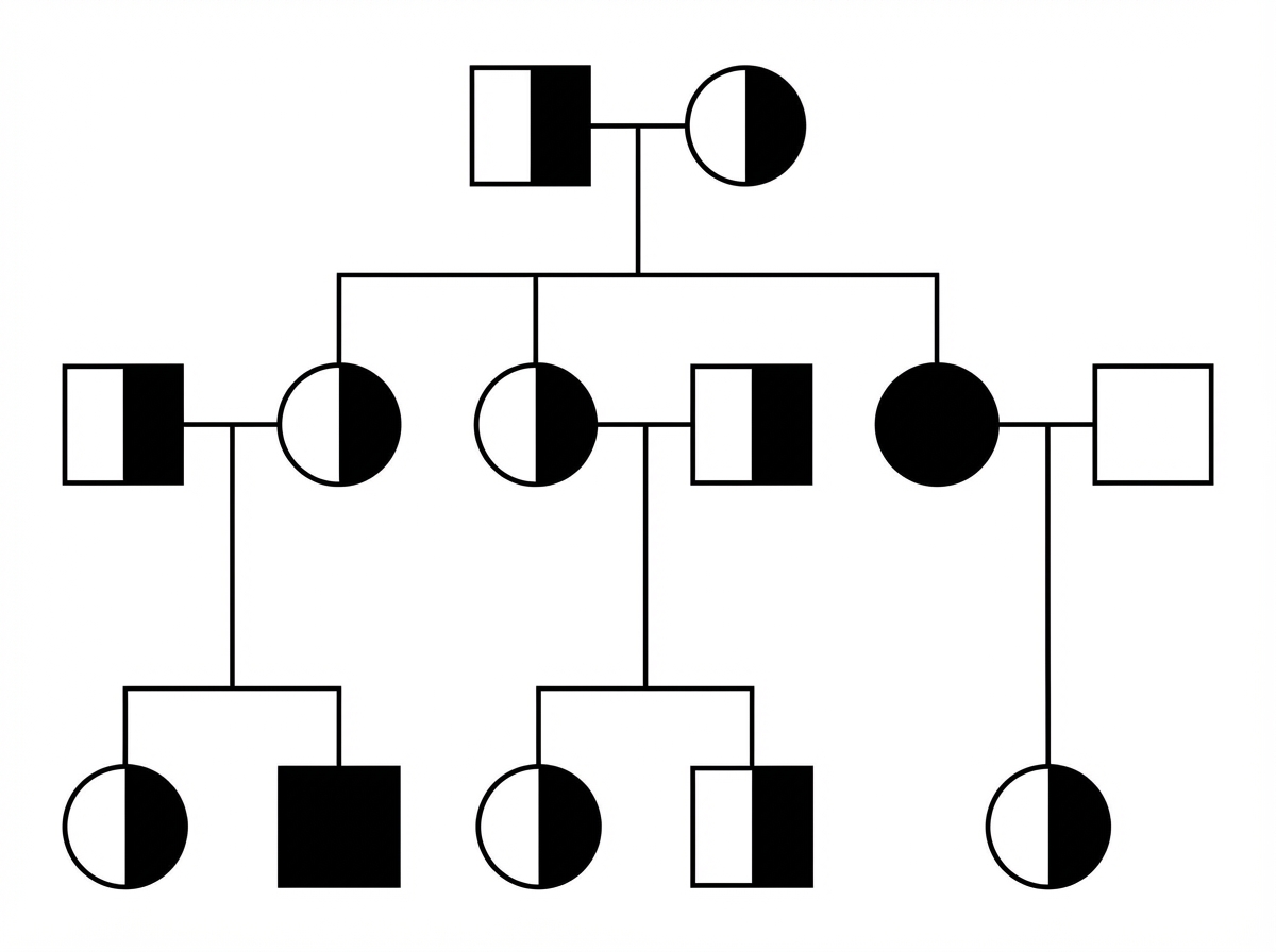
Examine the given pedigree chart. Which one of the following diseases is the most likely for this situation?

Myotonic dystrophy is inherited on which chromosome?
Unusual extensibility of the tongue is a characteristic feature of which of the following conditions?
Mental retardation is NOT a feature of which of the following mucopolysaccharidoses?
Duchenne's muscular dystrophy is inherited in which pattern?
Which condition has an autosomal dominant inheritance pattern?
Which single gene disorder does not follow Mendelian inheritance?
A 35-year-old nonsmoking male has been diagnosed with emphysema. His father died of emphysema at age 30, but he smoked. His father also had cirrhosis and recurrent pancreatitis but did not drink alcohol. Which one of the following inheritance patterns typifies this disease process?
Adult Polycystic kidney disease is inherited by which mode?
Explanation: Myotonic Dystrophy Type 1 (Steinert’s disease) is an autosomal dominant multisystemic disorder caused by a **CTG trinucleotide repeat expansion** in the DMPK gene on chromosome 19. It is characterized by "anticipation" and affects both skeletal muscle and various organ systems. **Why "Enlarged testis" is the correct answer:** In Myotonic Dystrophy, the endocrine system is significantly affected. Instead of enlargement, patients typically develop **primary testicular atrophy**, leading to hypogonadism, reduced fertility, and low testosterone levels. Therefore, "Enlarged testis" is clinically incorrect and is the right choice for this "except" type question. **Analysis of incorrect options:** * **Cardiac defects:** These are a major cause of morbidity. Patients frequently develop **conduction system abnormalities** (e.g., AV blocks, arrhythmias) and cardiomyopathy. * **Cataract:** A hallmark feature, often presenting as "Christmas tree cataracts" (iridescent subcapsular opacities) early in the disease. * **Frontal baldness:** A classic phenotypic feature in males, contributing to the characteristic "hatchet-faced" appearance (along with ptosis and wasting of temporalis/masseter muscles). **High-Yield Clinical Pearls for NEET-PG:** * **Grip Myotonia:** Difficulty releasing the hand after a handshake is a classic sign. * **Percussion Myotonia:** Elicited by tapping the thenar eminence. * **Other features:** Insulin resistance (Diabetes), sleep apnea, and intellectual disability (in congenital forms). * **Diagnosis:** Genetic testing for CTG repeats is the gold standard. EMG shows "dive bomber" potentials.
Explanation: ***Beta-thalassemia*** - The pedigree shows an **autosomal recessive** inheritance pattern with **carrier parents** who are unaffected but have affected children of both sexes. - **Generation skipping** is typical, as both parents must be carriers for children to be affected, matching beta-thalassemia transmission. *Neurofibromatosis* - Follows an **autosomal dominant** pattern where affected individuals appear in every generation without skipping. - At least one parent would be affected if a child has the condition, which contradicts the pedigree pattern. *Colour blindness* - Shows **X-linked recessive** inheritance, primarily affecting **males** with minimal female involvement. - Affected males cannot pass the condition to their sons, only to carrier daughters, inconsistent with this pedigree. *Vitamin D resistant rickets* - Demonstrates **X-linked dominant** inheritance where affected fathers pass the condition to **all daughters** but no sons. - The pedigree pattern doesn't match this specific father-to-daughter transmission pattern.
Explanation: **Explanation:** **Myotonic Dystrophy Type 1 (DM1)**, also known as Steinert’s disease, is the most common adult-onset muscular dystrophy. It is an **Autosomal Dominant** multisystem disorder caused by an unstable expansion of a **CTG trinucleotide repeat** in the 3' untranslated region of the **DMPK (Dystrophia Myotonica Protein Kinase) gene**, which is located on **Chromosome 19q13.3**. **Analysis of Options:** * **Option C (19):** This is the correct location for the DMPK gene (DM1). Note that Myotonic Dystrophy Type 2 (PROMM) is rarer and involves a CCTG expansion on Chromosome 3. * **Option A (21):** Chromosome 21 is primarily associated with Down Syndrome (Trisomy 21) and the APP gene related to early-onset Alzheimer’s. * **Option B (20):** Chromosome 20 is associated with conditions like Alagille syndrome and Creutzfeldt-Jakob disease (PRNP gene), but not myotonic dystrophy. * **Option D (24):** Humans only have 23 pairs of chromosomes (46 total). There is no "Chromosome 24" in the human genome. **High-Yield NEET-PG Pearls:** 1. **Anticipation:** The disease becomes more severe and has an earlier onset in successive generations due to the expansion of CTG repeats (especially via maternal transmission). 2. **Clinical Features:** "Hatchet facies" (temporalis wasting), frontal balding, cataracts, ptosis, and **myotonia** (delayed relaxation of muscles after contraction, e.g., difficulty releasing a handshake). 3. **Systemic Involvement:** Cardiac conduction defects (most common cause of sudden death), insulin resistance, and testicular atrophy. 4. **Diagnosis:** Genetic testing (PCR/Southern Blot) is the gold standard. EMG shows characteristic "dive bomber" discharges.
Explanation: The correct answer is **Ehlers-Danlos syndrome (EDS)**. This condition is a heterogeneous group of inherited connective tissue disorders characterized by defects in **collagen synthesis** (specifically Type I, III, or V, depending on the subtype) [1]. The hallmark of EDS is generalized joint hypermobility and skin hyperextensibility [1]. In the oral cavity, this connective tissue laxity manifests as the **Gorlin sign**, which is the ability to touch the tip of the nose with the tongue. This occurs in approximately 50% of EDS patients due to the unusual extensibility of the lingual frenulum and the tongue itself. **Analysis of Incorrect Options:** * **A. Epidermolysis bullosa:** A group of genetic conditions causing fragile, blistering skin. While it involves oral mucosal blistering and scarring (microstomia), it does not cause tongue hyperextensibility. * **B. Syphilis:** Tertiary syphilis may present with "interstitial glossitis" (leukoplakia and atrophy), but it typically leads to a firm, fibrotic tongue rather than an extensible one. * **C. Darier-White disease:** A keratinization disorder (keratosis follicularis) characterized by greasy hyperkeratotic papules. Oral involvement presents as "cobblestone" papules on the palate, not tongue laxity. **High-Yield Clinical Pearls for NEET-PG:** * **Gorlin Sign:** Pathognomonic for EDS (tongue to nose). * **Beighton Score:** Used to quantify joint hypermobility in EDS. * **Vascular EDS (Type IV):** Caused by a defect in **Type III collagen** (COL3A1); it is the most severe form due to the risk of arterial or organ rupture [1]. * **Other features:** "Cigarette paper" or "papyraceous" scars, easy bruising, and mitral valve prolapse (MVP).
Explanation: The correct answer is **D. Morquio syndrome (MPS IV)**. (Note: The provided key in the prompt incorrectly marks Sanfilippo; however, clinically and for NEET-PG, Morquio is the classic MPS characterized by **normal intelligence**). **1. Why Morquio Syndrome (MPS IV) is the correct answer:** In Mucopolysaccharisoses (MPS), mental retardation occurs when there is an accumulation of heparan sulfate in the CNS. Morquio syndrome is caused by a deficiency of *Galactose-6-sulfatase* (Type A) or *Beta-galactosidase* (Type B), leading to the accumulation of **Keratan sulfate** [1]. Since Keratan sulfate is not a primary component of the brain, patients with Morquio syndrome typically have **normal intelligence**. They are characterized by severe skeletal dysplasia (spondyloepiphyseal dysplasia) and ligamentous laxity. **2. Why the other options are incorrect:** * **Hurler Syndrome (MPS I):** This is the most severe form. It involves the accumulation of dermatan and heparan sulfate, leading to progressive developmental delay and significant cognitive impairment [1]. * **Hunter Syndrome (MPS II):** This is X-linked recessive. Like Hurler, it involves heparan sulfate accumulation, leading to mental retardation [1]. * **Sanfilippo Syndrome (MPS III):** This syndrome is unique because it presents with **severe CNS involvement** and behavioral disturbances, while the physical (somatic) features are relatively mild. Mental retardation is a hallmark feature [1]. **High-Yield Clinical Pearls for NEET-PG:** * **Normal Intelligence:** Morquio (MPS IV) and Scheie Syndrome (MPS IS) [1]. * **No Corneal Clouding:** Hunter Syndrome (MPS II) – "The Hunter needs clear eyes to see the target." * **X-linked Recessive:** Hunter Syndrome (All other MPS are Autosomal Recessive). * **Diagnosis:** Initial screening via urinary GAGs; confirmed by enzyme assay or genetic testing.
Explanation: **Explanation:** **Duchenne Muscular Dystrophy (DMD)** is caused by a mutation in the **DMD gene** located on the short arm of the **X chromosome (Xp21)**. This gene encodes for **dystrophin**, a critical protein that links the cytoskeleton of a muscle fiber to the surrounding extracellular matrix. 1. **Why X-linked Recessive (Correct):** DMD follows an X-linked recessive pattern because the gene is located on the X chromosome [1]. Since males have only one X chromosome (XY), a single mutated copy results in the disease. Females (XX) are typically asymptomatic carriers because their second, healthy X chromosome produces sufficient dystrophin. 2. **Why other options are incorrect:** * **X-linked Dominant:** In this pattern, both males and females would be affected if they carry just one copy of the mutation (e.g., Alport Syndrome). In DMD, females are generally spared from severe clinical symptoms. * **Autosomal Dominant/Recessive:** These involve mutations on non-sex chromosomes (1-22). DMD is specifically linked to the sex-determining X chromosome. **High-Yield Clinical Pearls for NEET-PG:** * **The Gene:** The DMD gene is the **largest known human gene**, making it highly susceptible to spontaneous mutations (30% of cases are *de novo*). * **Clinical Signs:** Look for **Gower’s sign** (using hands to "climb up" the legs to stand) and **pseudohypertrophy of the calves** (fatty replacement of muscle). * **Diagnosis:** Initial screen shows highly elevated **Creatine Kinase (CK)** levels. Gold standard is **Genetic Testing** (MLPA); Muscle biopsy shows absent dystrophin. * **Becker Muscular Dystrophy (BMD):** Also X-linked recessive but milder, caused by *in-frame* mutations resulting in truncated (partially functional) dystrophin.
Explanation: The correct answer is **Diamond-Blackfan Anaemia (DBA)**. This condition is a congenital pure red cell aplasia characterized by macrocytic anemia, reticulocytopenia, and a selective decrease in erythroid precursors in the bone marrow. **1. Why Diamond-Blackfan Anaemia is correct:** DBA is primarily inherited in an **Autosomal Dominant (AD)** pattern. It is caused by mutations in genes encoding ribosomal proteins (most commonly **RPS19**), leading to "ribosomal stress" and p53-mediated apoptosis of erythroid progenitor cells. Approximately 25% of patients also present with physical anomalies (triphalangeal thumbs, craniofacial defects). **2. Why the other options are incorrect:** * **Schwachman-Diamond Syndrome (SDS):** This is an **Autosomal Recessive (AR)** disorder characterized by exocrine pancreatic insufficiency and bone marrow failure (neutropenia). * **Dyskeratosis Congenita:** This is genetically heterogeneous but most commonly follows an **X-linked Recessive** pattern (DKC1 gene). While AD and AR forms exist, it is classically taught as X-linked in the context of the "triad" (nail dystrophy, leukoplakia, hyperpigmentation). * **Congenital Amegakaryocytic Thrombocytopenia (CAMT):** This is an **Autosomal Recessive (AR)** disorder caused by a mutation in the *MPL* gene (thrombopoietin receptor), leading to severe isolated thrombocytopenia at birth. **Clinical Pearls for NEET-PG:** * **DBA vs. TEC:** Transient Erythroblastopenia of Childhood (TEC) is an acquired mimic of DBA but occurs in older children (>1 year) and has no physical anomalies. * **Thumb Anomalies:** Both DBA and Fanconi Anemia (AR) feature thumb deformities; however, DBA is a pure red cell aplasia, while Fanconi is a pancytopenia. * **Biochemical Marker:** Elevated **Erythrocyte Adenosine Deaminase (eADA)** levels are a high-yield diagnostic marker for DBA.
Explanation: **Explanation:** The correct answer is **Fragile X syndrome** because it is a **Trinucleotide Repeat Expansion** disorder, which follows **Non-Mendelian inheritance**. ### 1. Why Fragile X syndrome is correct: Fragile X syndrome is caused by the expansion of **CGG repeats** in the *FMR1* gene on the X chromosome. Unlike Mendelian traits, which remain stable across generations, these repeats are unstable and can expand during gametogenesis. This leads to **Genetic Anticipation** (the disease becomes more severe or appears earlier in successive generations), a hallmark of non-Mendelian inheritance. ### 2. Why the other options are incorrect: * **Sickle cell anemia (A):** This is a classic **Autosomal Recessive** disorder caused by a point mutation in the HBB gene. It follows strict Mendelian laws of segregation and independent assortment. * **Down syndrome (B):** This is a **chromosomal numerical aberration** (Trisomy 21), usually due to meiotic non-disjunction. It is not a single-gene Mendelian disorder, but it is also not the answer here as the question asks for a "single gene disorder" that deviates from Mendelian patterns [1]. * **Retinoblastoma (D):** This follows **Autosomal Dominant** inheritance (though it requires a "second hit" at the cellular level, the pedigree remains Mendelian). ### NEET-PG High-Yield Pearls: * **Non-Mendelian Patterns:** Include Trinucleotide repeats (Fragile X, Huntington’s), Mitochondrial inheritance (LHON), Imprinting (Prader-Willi/Angelman), and Mosaicism. * **Fragile X Clinical Triad:** Intellectual disability, large ears, and macro-orchidism (post-pubertal). * **Southern Blot:** This is the gold standard for diagnosing Fragile X to determine the number of CGG repeats. * **Anticipation:** Always suspect a trinucleotide repeat disorder if a pedigree shows increasing severity in younger generations.
Explanation: The clinical presentation of early-onset emphysema in a non-smoker, combined with a family history of cirrhosis and pancreatitis, is classic for **Alpha-1 Antitrypsin (AAT) Deficiency**. **Why Codominant is Correct:** The inheritance of AAT deficiency is characterized by **codominance**. The *SERPINA1* gene has several alleles (M, S, and Z). In a codominant pattern, both alleles in a heterozygote are expressed equally, contributing to the overall phenotype. * **PiMM:** Normal levels of AAT. * **PiMZ:** Heterozygous; intermediate levels (increased risk if smoking). * **PiZZ:** Homozygous; severely low levels, leading to panacinar emphysema and liver cirrhosis (due to misfolding and accumulation of the protein in hepatocytes) [2]. **Why Incorrect Options are Wrong:** * **Autosomal Dominant:** In AD, one mutant allele masks the normal one. Here, the normal allele (M) still produces functional protein, so the phenotype depends on the specific combination of both alleles [1]. * **Incomplete Dominance:** This would imply a "blending" of traits where the phenotype is an intermediate between the two parents. In AAT deficiency, both gene products are distinctly present and measurable. * **Autosomal Recessive:** While the *disease* often manifests when two abnormal alleles are present (like ZZ), the *inheritance pattern* of the alleles themselves is codominant because both alleles contribute to the serum protein level. **High-Yield Clinical Pearls for NEET-PG:** * **Pathology:** Emphysema in AAT deficiency is **Panacinar** (lower lobes), whereas smoking-related emphysema is **Centriacinar** (upper lobes). * **Liver Involvement:** PAS-positive, diastase-resistant globules in hepatocytes are pathognomonic [1]. * **Diagnosis:** Initial screening is via serum AAT levels; gold standard is phenotyping (isoelectric focusing). * **Associated Condition:** Panniculitis (necrotizing skin lesions) is a rare but high-yield association.
Explanation: **Autosomal Dominant Polycystic Kidney Disease (ADPKD)** is the most common hereditary kidney disease [1]. It is inherited in an **Autosomal Dominant** pattern, meaning a single copy of the mutated gene from one parent is sufficient to cause the disease [1]. * **Why Option A is correct:** ADPKD is primarily caused by mutations in the **PKD1** gene (85% of cases, located on Chromosome 16) or the **PKD2** gene (15% of cases, located on Chromosome 4) [1]. It typically presents in the 3rd to 5th decade of life with bilateral renal cysts, hypertension, and hematuria [1]. * **Why Options B, C, and D are incorrect:** * **Autosomal Recessive (ARPKD):** This is a distinct, rarer entity caused by mutations in the *PKHD1* gene. It typically presents in infancy or childhood and is associated with congenital hepatic fibrosis. * **X-linked/Mitochondrial:** These modes of inheritance are not associated with the classic presentation of polycystic kidney disease. **High-Yield Clinical Pearls for NEET-PG:** 1. **Extra-renal manifestations:** The most common is **Liver cysts**. The most life-threatening is **Berry Aneurysms** (Circle of Willis), which can lead to subarachnoid hemorrhage. Other features include mitral valve prolapse and diverticulosis. 2. **Diagnosis:** Ultrasonography is the primary screening tool. Diagnosis is based on the number of cysts relative to the patient's age (Ravine criteria). 3. **Prognosis:** PKD1 mutations progress to End-Stage Renal Disease (ESRD) earlier (mean age ~54) compared to PKD2 (mean age ~74) [1]. 4. **Treatment:** Tolvaptan (a V2-receptor antagonist) is used to slow the progression of cyst growth and renal decline.
Principles of Medical Genetics
Practice Questions
Genetic Testing and Counseling
Practice Questions
Single Gene Disorders
Practice Questions
Chromosomal Disorders
Practice Questions
Mitochondrial Diseases
Practice Questions
Pharmacogenomics
Practice Questions
Cancer Genetics
Practice Questions
Genetics of Common Diseases
Practice Questions
Epigenetics and Disease
Practice Questions
Genetic Basis of Developmental Disorders
Practice Questions
Ethical Issues in Medical Genetics
Practice Questions
Gene Therapy and Precision Medicine
Practice Questions
Get full access to all questions, explanations, and performance tracking.
Start For Free