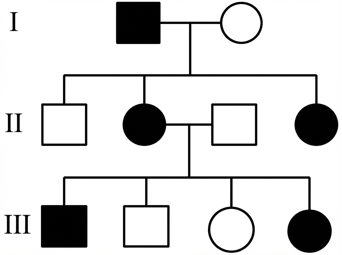
Which is the most likely inheritance pattern of the disease shown in the family pedigree?

Von Hippel-Lindau disease is associated with all the following except?
Phenotypic expression of a gene depending on the parent of origin is referred to as _______
Which of the following is the inheritance pattern of Incontinentia Pigmenti?
Mucociliary clearance is increased in cystic fibrosis by which of the following agents?
In patients with hypertrophic cardiomyopathy, in which gene are the maximum mutations found?
Which of the following is NOT an autosomal dominant disorder?
Which condition is Autosomal dominant?
Classic form of Alport syndrome is inherited as:
Which of the following statements about Hemochromatosis is true?
Explanation: ***X-Linked dominant type*** - **Affected fathers** pass the condition to **all daughters** but **no sons**, as fathers pass their X chromosome only to daughters. - **Affected females** appear in **every generation** with **vertical transmission**, distinguishing it from X-linked recessive patterns. *Autosomal recessive type* - Would show **horizontal transmission** with affected siblings but unaffected parents in typical cases. - Both **males and females** would be equally affected, and **consanguinity** would often be present in family history. *Autosomal dominant type* - Would show **equal transmission** to both male and female offspring from affected parents. - **Affected fathers** would pass the condition to approximately **50% of both sons and daughters**, not exclusively to daughters. *X-linked recessive type* - Would primarily affect **males** with **carrier females** who are typically unaffected. - Shows **knight's move pattern** with affected males related through unaffected carrier females, and **no male-to-male transmission**.
Explanation: **Explanation** Von Hippel-Lindau (VHL) disease is an autosomal dominant multisystem neoplastic syndrome caused by a mutation in the **VHL gene on chromosome 3p25**. The question asks which condition is *not* associated with VHL; however, based on clinical pathology, **Hemangioblastomas (Option B) are actually the hallmark lesion of VHL.** *Note: In the context of "Except" questions in medical exams, if all options are technically associated, the "correct" answer usually refers to the most common feature or a distractor. However, in standard medical literature, all four options listed are recognized components of VHL.* **Breakdown of Associations:** * **Hemangioblastomas (Option B):** These are the most common manifestation, typically occurring in the cerebellum, retina, and spinal cord [2]. Retinal hemangioblastomas (angiomas) are often the presenting sign. * **Pheochromocytoma (Option C):** VHL Type 2 is specifically characterized by a high risk of pheochromocytomas, which are often bilateral and multifocal. * **Islet Cell Tumors (Option D):** Pancreatic involvement is common, ranging from simple cysts to Pancreatic Neuroendocrine Tumors (PNETs/Islet cell tumors) [1]. * **Endolymphatic Sac Tumors (Option A):** These are highly specific (though less common) vascular tumors of the inner ear that can lead to hearing loss and vertigo in VHL patients. **High-Yield Clinical Pearls for NEET-PG:** * **VHL Gene Function:** It encodes a protein that part of an E3 ubiquitin ligase complex which degrades **Hypoxia-Inducible Factor (HIF)**. Loss of VHL leads to abnormal accumulation of HIF, causing overexpression of VEGF and Erythropoietin [2]. * **Renal Cell Carcinoma (RCC):** VHL is the most common cause of inherited **Clear Cell RCC**, which is often bilateral and occurs at a young age [2]. * **Mnemonic (HIPPEL):** **H**emangioblastomas, **I**slet cell tumors, **P**heochromocytoma, **P**ancreatic cysts, **E**ndolymphatic sac tumors, **L**eural (Renal) Cell Carcinoma.
Explanation: **Explanation:** **Genomic Imprinting** is an epigenetic process where certain genes are expressed in a parent-of-origin-specific manner. While most autosomal genes are expressed from both alleles, imprinted genes are "silenced" (usually via DNA methylation) in either the egg or the sperm. Therefore, the phenotype of the offspring depends entirely on which parent transmitted the active allele [1]. **Analysis of Incorrect Options:** * **B. Mosaicism:** Refers to the presence of two or more populations of cells with different genotypes in one individual, derived from a single zygote (e.g., Turner syndrome mosaicism 45,X/46,XX). * **C. Anticipation:** A phenomenon where a genetic disorder becomes more severe or appears at an earlier age in succeeding generations. This is typically seen in **Trinucleotide Repeat Disorders** (e.g., Huntington’s disease, Fragile X). * **D. Nonpenetrance:** Occurs when an individual carries a dominant disease-causing mutation but does not exhibit any clinical symptoms of the disease. **High-Yield Clinical Pearls for NEET-PG:** * **Prader-Willi Syndrome:** Deletion of the **paternal** copy of chromosome 15q11-q13 (Maternal imprinting). Features: Hyperphagia, obesity, and hypogonadism. * **Angelman Syndrome:** Deletion of the **maternal** copy of the same region (Paternal imprinting) [1]. Features: Inappropriate laughter ("Happy Puppet"), ataxia, and seizures. * **Uniparental Disomy (UPD):** When an individual receives two copies of a chromosome from one parent and none from the other; this is a common mechanism for imprinting disorders [1].
Explanation: **Explanation:** **Incontinentia Pigmenti (IP)**, also known as Bloch-Sulzberger syndrome, is a rare multi-system neurocutaneous disorder. The correct answer is **X-linked dominant (Option C)** because the disease is caused by a mutation in the **IKBKG gene** (formerly NEMO) located on the X chromosome. 1. **Why X-linked Dominant is correct:** In IP, the mutation is lethal in most hemizygous males (resulting in spontaneous abortion). It primarily affects females, who survive due to functional mosaicism resulting from **X-chromosome inactivation (Lyonization)** [1]. This explains the characteristic "Blaschko’s lines" distribution of skin lesions. 2. **Why other options are incorrect:** * **Autosomal Dominant/Recessive:** The gene is located on the X chromosome, not an autosome. * **X-linked Recessive:** IP does not follow a recessive pattern; a single mutated allele in females is sufficient to manifest the disease (dominant expression), while males typically do not survive to be "carriers" or affected patients. **High-Yield Clinical Pearls for NEET-PG:** * **Four Stages of Skin Lesions:** 1. Vesicular (Linear vesicles, birth to 4 months) 2. Verrucous (Hyperkeratotic) 3. Hyperpigmented (Swirls/Whirls of "Chinese-figure" pigmentation) 4. Atrophic/Hypopigmented (Scarring). * **Associated Findings:** Peg-shaped teeth (hypodontia), CNS abnormalities (seizures), and ocular defects (retinal neovascularization). * **Key Concept:** If a question mentions a "lethal mutation in males" with "swirl-like skin pigmentation in a female child," always think of Incontinentia Pigmenti.
Explanation: ### Explanation **Correct Option: B (Amiloride)** **Mechanism and Concept:** In Cystic Fibrosis (CF), the defect in the CFTR protein leads to two major ionic imbalances in the airway epithelium: decreased chloride secretion and **increased sodium reabsorption** via the Epithelial Sodium Channel (ENaC) [1]. This excessive sodium uptake causes water to follow osmotically out of the airway lumen, leading to dehydrated, viscid mucus that impairs mucociliary clearance [1]. **Amiloride** is a potassium-sparing diuretic that acts as an **ENaC inhibitor**. When administered as an aerosol, it blocks the hyperactive sodium channels in the respiratory tract. By preventing excessive sodium (and subsequently water) reabsorption, it helps maintain airway surface liquid volume, thins the mucus, and thereby **increases mucociliary clearance**. **Analysis of Incorrect Options:** * **A. Mannitol:** While inhaled hypertonic saline and dry powder mannitol are used as osmotic agents to hydrate mucus, they do not directly inhibit the sodium channels responsible for the primary pathophysiology. In many standardized exams, Amiloride is the classic pharmacological answer for ENaC-mediated clearance improvement. * **C. Acetazolamide:** This is a carbonic anhydrase inhibitor used primarily for glaucoma, altitude sickness, and metabolic alkalosis [2]. It has no significant role in improving airway mucociliary clearance in CF. **High-Yield Clinical Pearls for NEET-PG:** * **CFTR Mutation:** Most common is **ΔF508** (Class II defect: protein misfolding and trafficking failure). * **Diagnosis:** Sweat chloride >60 mEq/L is the gold standard. * **Other Clearance Agents:** Recombinant human DNase (**Dornase alfa**) is used to digest extracellular DNA from neutrophils in CF sputum to reduce viscosity. * **Ivacaftor:** A CFTR "potentiator" used for specific gating mutations (Class III).
Explanation: Hypertrophic Cardiomyopathy (HCM) is an autosomal dominant genetic disorder characterized by unexplained left ventricular hypertrophy [1]. It is fundamentally a **disease of the sarcomere**, caused by mutations in genes encoding proteins of the cardiac contractile apparatus [1]. **1. Why Beta-myosin heavy chain (MYH7) is correct:** The two most common genes involved in HCM are **MYH7** (encoding Beta-myosin heavy chain) and **MYBPC3** (encoding Myosin-binding protein C) [1]. Historically and in most classic literature, **MYH7** is cited as the gene where the maximum number of mutations are identified, accounting for approximately 30-35% of all cases. It was the first gene linked to HCM and is associated with a higher risk of sudden cardiac death [1]. **2. Why the other options are incorrect:** * **Elastin:** Mutations in the elastin gene are associated with **Williams Syndrome** and supravalvular aortic stenosis, not HCM. * **Alpha-tropomyosin (TPM1):** While this is a sarcomeric gene involved in HCM, it accounts for a very small percentage of cases (<5%). * **Troponin T (TNNT2):** This gene accounts for about 5-10% of cases [1]. Notably, mutations here often cause mild hypertrophy but carry a high risk of sudden death [1]. **Clinical Pearls for NEET-PG:** * **Inheritance:** Autosomal Dominant with variable penetrance [1]. * **Most common cause of Sudden Cardiac Death (SCD)** in young athletes. * **Histology:** Characterized by **myocyte disarray** and interstitial fibrosis [1]. * **Murmur:** Harsh systolic ejection murmur that **increases** with Valsalva or standing (decreased preload) and **decreases** with squatting or handgrip (increased afterload/preload).
Explanation: **Explanation:** The correct answer is **Haemochromatosis**. Hereditary Haemochromatosis (specifically Type 1, involving the *HFE* gene) is classically inherited in an **Autosomal Recessive (AR)** pattern [1]. It is characterized by excessive intestinal iron absorption leading to iron overload in organs like the liver, heart, and pancreas. **Analysis of Options:** * **Haemochromatosis (Option C):** As an AR disorder, it requires two copies of the mutated gene (e.g., C282Y or H63D) for phenotypic expression [1]. It is a classic "exception" often tested because it is common in Caucasian populations but follows recessive inheritance. * **Myotonic Dystrophy (Option A):** This is the most common adult-onset muscular dystrophy and follows an **Autosomal Dominant (AD)** pattern. It is notable for "Anticipation" due to CTG trinucleotide repeat expansions. * **Von Willebrand Disease (Option B):** Most types (especially Type 1 and Type 2) are inherited in an **AD** fashion. It is the most common inherited bleeding disorder. * **Marfan’s Syndrome (Option D):** This is a classic **AD** connective tissue disorder caused by mutations in the *FBN1* gene on chromosome 15, leading to fibrillin-1 deficiency. **High-Yield Clinical Pearls for NEET-PG:** * **Mnemonic for AD disorders:** "**V**ery **P**owerful **M**any **M**en": **V**on Willebrand, **P**olycystic Kidney Disease (Adult), **M**yotonic Dystrophy, **M**arfan’s. * **Haemochromatosis Triad:** "Bronze Diabetes" (Hyperpigmentation, Diabetes Mellitus, and Cirrhosis). * **Key Distinction:** Most structural protein defects are AD (e.g., Marfan's), while most enzyme deficiencies are AR (e.g., Haemochromatosis, Wilson’s disease) [2].
Explanation: **Explanation:** **Correct Answer: D. Adult polycystic kidney disease (ADPKD)** Adult Polycystic Kidney Disease is a classic example of an **Autosomal Dominant (AD)** disorder [1]. It is primarily caused by mutations in the **PKD1** (Chromosome 16) or **PKD2** (Chromosome 4) genes [1]. In AD inheritance, only one copy of the mutated gene is required for the disease to manifest, and it typically appears in every generation (vertical transmission). **Analysis of Incorrect Options:** * **A. Hemophilia A:** This is an **X-linked Recessive** disorder caused by a deficiency of Factor VIII. It primarily affects males, while females are typically asymptomatic carriers. * **B. Duchenne muscular dystrophy (DMD):** This is also an **X-linked Recessive** condition. It involves a mutation in the *DMD* gene (the largest known human gene) on the X chromosome, leading to a lack of the protein dystrophin. * **C. Wilson's disease:** This is an **Autosomal Recessive** disorder of copper metabolism (ATP7B gene on Chromosome 13). Both parents must carry the mutated gene for the offspring to be affected. **High-Yield Clinical Pearls for NEET-PG:** * **ADPKD Associations:** Often associated with **Berry aneurysms** (Circle of Willis), hepatic cysts, and mitral valve prolapse (MVP). * **Mnemonic for AD Disorders:** "Very Powerful DOMINANT" – **V**on Willebrand, **P**olyposis (FAP), **D**ystrophia myotonica, **O**steogenesis imperfecta, **M**arfan, **I**ntermittent porphyria, **N**eurofibromatosis, **A**chondroplasia, **N**oonan, **T**uberous sclerosis. * **Rule of Thumb:** Most structural protein defects are Autosomal Dominant, while most enzyme deficiencies are Autosomal Recessive.
Explanation: Alport syndrome is a primary glomerular disease caused by mutations in the genes encoding **Type IV collagen**, which is a critical structural component of the glomerular basement membrane (GBM), cochlea, and lens. **1. Why X-linked is Correct:** The "classic" and most common form of Alport syndrome (accounting for approximately **80-85% of cases**) is inherited in an **X-linked dominant** fashion. This is due to mutations in the **COL4A5 gene** located on the X chromosome. Because it is X-linked, males are typically more severely affected, often progressing to end-stage renal disease (ESRD), while females (heterozygotes) may have a more variable clinical course ranging from isolated hematuria to renal failure. **2. Why other options are Incorrect:** * **Autosomal Recessive (AR):** This accounts for about 15% of cases and is caused by mutations in *COL4A3* or *COL4A4*. It is seen in clinical practice but is not the "classic" or most frequent form. * **Autosomal Dominant (AD):** This is the rarest form (approx. 5%) and usually presents with a slower progression to renal failure. * **Sporadic:** While de novo mutations can occur in the *COL4A5* gene, Alport syndrome is fundamentally a hereditary genetic disorder, not a sporadic one. **High-Yield Clinical Pearls for NEET-PG:** * **Clinical Triad:** Hereditary nephritis (hematuria), Sensorineural hearing loss (high frequency), and Ocular defects (**Anterior Lenticonus** is pathognomonic). * **Electron Microscopy (EM):** Characterized by a "Basket-weave appearance" due to irregular thinning and thickening of the GBM. * **Genetic Defect:** Mutation in alpha-5 chain of Type IV collagen (X-linked). * **Post-Transplant Complication:** Patients with Alport syndrome may develop **Goodpasture-like syndrome** (Anti-GBM disease) after a kidney transplant because their immune system recognizes the normal Type IV collagen in the graft as foreign.
Explanation: Explanation: Hereditary Hemochromatosis (HH) is a disorder of iron metabolism characterized by increased intestinal iron absorption, leading to iron deposition in various organs (liver, heart, pancreas, and joints). 1. Why Option D is Correct: The most common form of Hemochromatosis (Type 1) is caused by mutations in the HFE gene (most commonly C282Y) located on Chromosome 6 [2]. It is inherited as an autosomal recessive disorder. This means an individual must inherit two mutated alleles to be at risk for the phenotypic expression of the disease [2]. Both the C282Y and the H63D mutations can be identified by genetic testing, which is now in routine clinical use [1]. 2. Why the Other Options are Incorrect: * Option A: While phlebotomy is the treatment of choice and can normalize life expectancy if started early, it is not "curative" in a genetic sense [1]. The underlying genetic defect remains, and the patient requires lifelong monitoring and maintenance therapy [1]. * Option B: Although the gene frequency is equal in both sexes, the disease is more common in males (approx. 5:1 to 10:1 ratio). Females are "physiologically protected" due to iron loss during menstruation and pregnancy, delaying clinical presentation. * Option C: Hemochromatosis is a classic example of incomplete penetrance. Many individuals homozygous for the C282Y mutation never develop clinical symptoms or significant iron overload [1]. Clinical Pearls for NEET-PG: * Classic Triad (Bronze Diabetes): Cirrhosis, Diabetes Mellitus, and Skin Hyperpigmentation [2]. * Early Sign: Arthropathy (specifically involving the 2nd and 3rd metacarpophalangeal joints). * Diagnosis: Best initial test is Transferrin Saturation (>45%); Gold standard for diagnosis is HFE Gene Analysis [1]. * MRI: Used to quantify hepatic iron (shows low signal intensity on T2-weighted images) [1]. * Most common cause of death: Hepatocellular Carcinoma (HCC) [2].
Principles of Medical Genetics
Practice Questions
Genetic Testing and Counseling
Practice Questions
Single Gene Disorders
Practice Questions
Chromosomal Disorders
Practice Questions
Mitochondrial Diseases
Practice Questions
Pharmacogenomics
Practice Questions
Cancer Genetics
Practice Questions
Genetics of Common Diseases
Practice Questions
Epigenetics and Disease
Practice Questions
Genetic Basis of Developmental Disorders
Practice Questions
Ethical Issues in Medical Genetics
Practice Questions
Gene Therapy and Precision Medicine
Practice Questions
Get full access to all questions, explanations, and performance tracking.
Start For Free