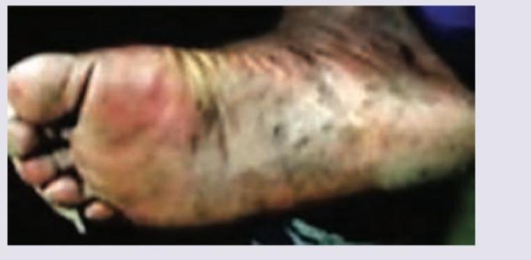
The following autopsy finding occurs due to:

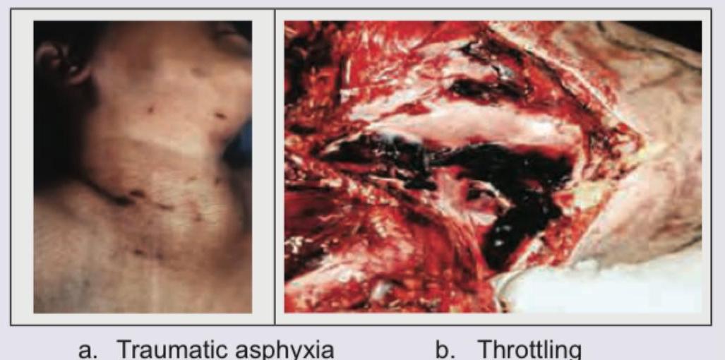
A person was brought dead to the casualty and autopsy was performed to find the cause of death. Based on the autopsy findings shown in the image, what is the diagnosis?

The following presentation is called:

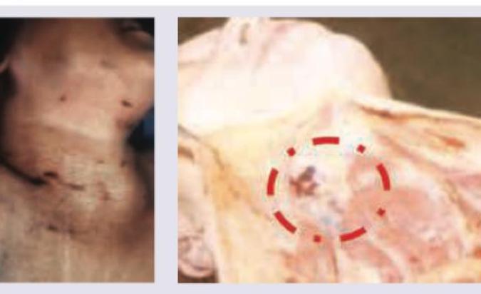
A female was found dead in her bedroom. The room was not locked from inside. Her blood alcohol value was found to be 350 mg/dL. The picture taken at the post mortem is shown below. The diagnosis is? (AIIMS Nov 2018, AIIMS Nov 2017)

The clinical signs of brain-stem death include all of the following except:
In autopsy, which organ is removed with liver?
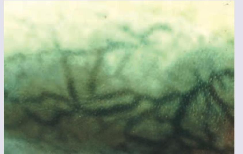
The component of vitreous humour that is most useful in determining time since death?
Certain obligations on the part of a doctor who undertakes a postmortem examination are the following, EXCEPT:
For autopsy, vitreous is preserved in:
Cardiac chambers are opened in autopsy in which order? i) Left atrium ii) Left ventricle iii) Right atrium iv) Right ventricle
Explanation: ***Sulfameth-haemoglobin*** - The image depicts **greenish discoloration** of tissues, which is characteristic of **post-mortem sulfhemoglobin formation** during putrefaction. - This occurs when **hydrogen sulfide** (produced by bacterial decomposition) reacts with hemoglobin to form sulfhemoglobin, causing the characteristic **green discoloration** seen in putrefying bodies. - This is a **normal post-mortem change** that typically begins in the right iliac fossa and spreads, appearing 24-48 hours after death in temperate climates. - The greenish hue is most prominent over the **abdomen** due to proximity to intestinal bacteria. *Hematin crystals* - **Hematin crystals** are formed from digested blood and appear as **dark brown to black, rhomboid crystals** microscopically, often indicating old hemorrhage. - They do not cause an overall greenish discoloration of the body, but rather represent localized microscopic findings. *Carboxy-haemoglobin* - **Carboxyhemoglobin** forms from carbon monoxide poisoning and causes a characteristic **cherry-red or pink discoloration** of tissues, blood, and livor mortis due to carbon monoxide binding to hemoglobin. - This is distinctly different from the greenish appearance associated with putrefaction. *Met-haemoglobin* - **Methemoglobinemia** results from oxidation of hemoglobin iron to the ferric state, causing a **chocolate-brown or grayish discoloration** of blood and tissues. - This appears as brownish discoloration, not the characteristic green hue of putrefactive changes.
Explanation: ***Hanging*** - The image shows **ligature marks on the neck**, consistent with hanging. Specifically, there is evidence of **fracture of the hyoid bone** and possibly other laryngeal cartilages (circled area), which are common findings in hanging, especially **judicial hanging** or cases with a significant drop. - The presence of the ligature mark **above the thyroid cartilage** and extending upwards towards the mastoid process is characteristic of hanging, creating an **oblique, upward-sloping** V-shaped or inverted U-shaped groove. - In hanging, the body weight acts as the constricting force, and the mark is typically **incomplete** on the side opposite to the suspension point. *Throttling* - Throttling involves **manual compression of the neck** by human hands. It typically leaves **finger marks** and **bruises** on the neck, often irregular and distributed over a wider area. - While hyoid bone fractures can occur in throttling, the distinct, continuous **ligature mark** and the specific position of injuries seen in the image are less typical for manual strangulation. *Strangulation (general)* - Strangulation refers to compression of the neck by a constricting force, which can be manual (throttling), ligature (ligature strangulation), or by hanging. Without further context, this term is too broad and non-specific. *Ligature strangulation* - Ligature strangulation differs from hanging in that the constricting force is applied around the neck by a ligature (rope, wire, cloth), but the body is **not suspended**. - This typically produces **horizontal or transverse ligature marks** that are more uniform and circumferential, unlike the **oblique, upward-sloping marks** characteristic of hanging. - The ligature mark in strangulation is usually **complete** around the neck and positioned lower, often at the level of the thyroid cartilage.
Explanation: ***Mycetoma*** - The image displays a foot with **swelling, nodule formation, and draining sinuses** with visible grains, which is characteristic of mycetoma. - Mycetoma is a **chronic, progressive granulomatous disease** of the skin, subcutaneous tissue, fascia, and bone, often affecting the foot. *Black foot disease* - This is a form of **peripheral vascular disease** caused by **chronic arsenic poisoning**, typically leading to **gangrene** and eventually amputation. - The image does not show the typical features of **ischemia or gangrene** associated with Black foot disease. *Myrmecia* - This is a type of **wart or verruca** that is specifically characterized by a **plantar wart with a central core of black dots** (thrombosed capillaries). - The image shows a more widespread inflammatory process with nodules and sinuses, not a localized wart. *Tinea pedis* - Also known as **athlete's foot**, this is a **superficial fungal infection** of the skin of the foot, typically presenting with **scaling, itching, redness, and sometimes blisters**. - The clinical presentation in the image, with deep-seated nodules and draining sinuses, is not consistent with a superficial fungal infection.
Explanation: ***Throttling*** - The autopsy image shows extensive **internal hemorrhage** and disruption of neck structures, consistent with significant compressive force applied to the neck by hands, as seen in **throttling**. - **Throttling** (manual strangulation) causes deep internal injuries including fractured **hyoid bone**, damaged **thyroid cartilage**, and **strap muscle hemorrhage**, even when external marks may be minimal or absent. - The high blood alcohol level (350 mg/dL - severe intoxication) would have impaired her ability to resist, and the unlocked room suggests **homicidal** intent rather than suicide. - Key autopsy findings: **deep neck muscle hemorrhage**, **laryngeal fractures**, and **torn blood vessels** without a ligature pattern. *Traumatic asphyxia* - Traumatic asphyxia results from severe **chest/thoracic compression** leading to acute venous congestion in the head and neck region. - Classical signs include **petechial hemorrhages** on face and conjunctivae, **cyanosis** above compression level, and relatively **intact neck structures** on autopsy. - The severe internal neck damage shown in the autopsy image is **not characteristic** of traumatic asphyxia, which primarily affects superficial vessels due to back-pressure, not deep structural injury. *Café coronary* - **Café coronary** is sudden death from **food bolus aspiration** causing airway obstruction, commonly occurring in intoxicated individuals who cannot protect their airway. - Autopsy findings would show an **obstructing food bolus in the larynx/trachea** without the extensive neck trauma and hemorrhage depicted in the image. - No manual strangulation injuries would be present. *Bansdola* - **Bansdola** is a traditional method of strangulation using a **bamboo stick or rod** twisted across the neck with a rope, used historically as torture or execution. - It causes a characteristic **linear ligature mark** with underlying soft tissue injury in a horizontal pattern across the neck. - The autopsy findings in the image show **diffuse manual strangulation injury** rather than the specific linear pattern of ligature strangulation seen in Bansdola.
Explanation: **Profound hypotension** - While patients with brainstem death can be hemodynamically unstable, **profound hypotension** itself is not a defining diagnostic criterion for brainstem death. It is often a consequence of brainstem injury, but not a direct sign indicating loss of brainstem function. - The criteria for brainstem death focus on the irreversible cessation of all brainstem reflexes and the capacity for spontaneous respiration, not peripheral circulatory status. *Absence of spontaneous respiration* - The **apnea test**, demonstrating no respiratory efforts despite adequate CO2 stimulation, is a critical component of brainstem death diagnosis, indicating irreversible loss of brainstem respiratory centers. - This sign confirms the total and irreversible cessation of the brainstem's ability to maintain vital functions. *Absent oculo-vestibular reflex* - The absence of the **oculo-vestibular reflex** (cold caloric test) indicates irreversible damage to the brainstem nuclei responsible for eye movements and vestibular function. - This is a key diagnostic criterion demonstrating the loss of specific brainstem reflexes. *Absence of pharyngeal reflex* - The absence of the **pharyngeal reflex** (gag reflex) signifies the loss of cranial nerve IX and X function, which are mediated by the brainstem. - This confirms the irreversible cessation of a vital protective reflex regulated by the brainstem.
Explanation: ***Spleen*** - In autopsy, the **spleen is routinely removed along with the liver** as part of standard dissection protocols (Virchow's method and variations). - This is done to examine the **portal venous system**, assess the **hepatosplenic circulation**, and evaluate pathologies affecting both organs such as **portal hypertension**, **congestive splenomegaly**, or **hepatic cirrhosis**. - The anatomical proximity and shared vascular connections make their removal together both practical and diagnostically valuable. *Kidney* - The **kidneys** are typically removed separately or as part of the **posterior abdominal block** after the liver and spleen have been removed. - While kidneys may be examined in relation to the abdominal vasculature, they are not removed in direct continuity with the liver block in standard autopsy technique. - Their retroperitoneal location makes separate dissection more appropriate. *Stomach* - The **stomach** is removed as part of the **gastrointestinal tract block**, which includes the esophagus, duodenum, and often the pancreas. - Though anatomically adjacent to the liver, it is not part of the hepatosplenic block. - Its examination focuses on mucosal pathology, ulcers, tumors, and gastric contents rather than hepatic circulation. *Brain* - The **brain** is removed as a completely separate organ through **craniotomy** after reflection of the scalp. - It is housed within the cranium and has no anatomical or dissection relationship with abdominal organs. - Brain removal follows examination of the cranial cavity, meninges, and cerebral vessels.
Explanation: ***Potassium*** - **Potassium** concentration in the **vitreous humor** increases steadily after death due to the breakdown of cell membranes and pumps, making it a reliable biochemical marker for estimating **time since death (postmortem interval)**. - Its rate of diffusion from cells into the vitreous is relatively constant and less affected by external factors compared to other electrolytes. *Sodium* - **Sodium** levels in the vitreous humor are generally stable postmortem for a period but can fluctuate, making it less reliable than potassium for precise **postmortem interval (PMI)** estimation. - Its concentration is influenced by osmolality changes and is not a primary indicator of linear cellular breakdown over time. *Magnesium* - While present in the vitreous, **magnesium** concentration changes postmortem are less predictable and less extensively studied compared to potassium, making it less commonly used for **PMI** determination. - Its release from cells is not as linear or substantial as potassium's over the typical postmortem period. *Calcium* - **Calcium** levels in the vitreous humor show some postmortem changes, but these increases are often less consistent and slower than potassium, making it a less accurate marker for **PMI** estimation. - Its involvement in cellular processes and potential postmortem precipitation can introduce variability, limiting its utility.
Explanation: ***He must keep the police informed about the findings*** - This is **NOT a formal obligation** of the doctor conducting a postmortem examination. - The doctor's primary duty is to conduct a thorough, objective examination and prepare a **formal postmortem report** that is submitted to the authority who requisitioned the examination (magistrate/police as per CrPC Section 174). - While findings may eventually reach the police through the official report, there is **no obligation to informally update or keep police informed** during the examination process. - The doctor's role is that of an **independent expert witness** to the court, not an investigative assistant to the police. - Maintaining independence and objectivity requires the doctor to document findings formally rather than providing ongoing informal updates to investigating officers. *Routinely record all positive findings and important negative ones* - This IS a **fundamental obligation** for any doctor performing a postmortem examination. - Both positive findings (pathological changes, injuries) and significant negative findings (absence of expected pathology) must be documented to provide a comprehensive and accurate record. - This meticulous documentation ensures the **integrity, reliability, and legal validity** of the postmortem examination and its conclusions. *The examination should be meticulous and complete* - This IS a **professional, ethical, and legal obligation** for any doctor undertaking a postmortem examination. - A systematic and thorough examination of all body systems is essential to accurately determine the cause of death and identify all relevant findings. - Incomplete examinations can lead to **missed diagnoses and miscarriage of justice** in medico-legal cases. *He must preserve viscera and send for toxicology examination in case of poisoning* - This IS a **crucial obligation** when poisoning is suspected or cannot be ruled out based on the postmortem findings. - Relevant viscera (liver, kidney, stomach contents) and bodily fluids (blood, urine) must be preserved in appropriate containers for subsequent toxicological analysis. - This step is **essential to confirm or exclude toxicological involvement** in the death and is a standard protocol in medico-legal postmortem examinations as per established guidelines.
Explanation: ***Fluoride (Sodium fluoride)*** - **Sodium fluoride** is the preferred preservative for vitreous humor samples collected during autopsy, especially when testing for **alcohol** and **glucose** levels. - It acts as an **enzyme inhibitor**, preventing post-mortem metabolism of glucose and fermentation of alcohol by microorganisms, thus maintaining the sample's integrity. *Phenol* - **Phenol** is an antiseptic and disinfectant often used in tissue preservation for histological examination, but it is not typically used for vitreous humor to preserve metabolites like glucose or alcohol. - It can interfere with some analytical methods used for vitreous analysis. *Sulphuric acid* - **Sulphuric acid** is a strong acid that would cause significant denaturation of proteins and alteration of the chemical composition of vitreous humor. - It is not suitable for preserving biological samples for most analyses, especially for fragile metabolites such as glucose. *Xylol* - **Xylol** (xylene) is a common solvent used in histology for clearing tissues (removing alcohol) before paraffin embedding, making it unsuitable for direct preservation of body fluids like vitreous humor. - It would dissolve cellular components and denature proteins, rendering the sample useless for chemical analysis.
Explanation: ***iii - iv - i - ii*** - This order (right atrium, right ventricle, left atrium, left ventricle) is the **standard and systematic approach** for opening cardiac chambers during an autopsy. - It allows for a **logical flow of examination**, mirroring the direction of blood flow through the heart and minimizing disruption to adjacent structures. *i - iii - ii - iv* - Opening the **left atrium first** before the right side of the heart is not a standard or systematic approach in cardiac dissection. - This order would disrupt the **natural progression** of blood flow examination and could complicate subsequent steps. *i - ii - iii - iv* - This order begins with the **left atrium and left ventricle** before moving to the right side, which is not the conventional method. - Starting with the left side first can make it difficult to properly assess the **overall cardiac structure** and connectivity in a logical sequence. *ii - iv - iii - i* - This order begins with the **ventricles first** which can make it challenging to properly assess the integrity of the atria and their connections with the great vessels. - It deviates from the standard practice of opening the atria before the ventricles to maintain a **systematic and thorough examination**.
Objectives of Medicolegal Autopsy
Practice Questions
Autopsy Procedures
Practice Questions
External Examination
Practice Questions
Internal Examination
Practice Questions
Special Autopsy Techniques
Practice Questions
Organ Retention and Disposal
Practice Questions
Collection of Toxicological Samples
Practice Questions
Autopsy Report Writing
Practice Questions
Histopathology in Autopsies
Practice Questions
Microbiology in Autopsies
Practice Questions
Radiology in Autopsies
Practice Questions
Limitations and Artifacts
Practice Questions
Get full access to all questions, explanations, and performance tracking.
Start For Free