
Fungal Skin Infections — MCQs

On this page
A child presents with multiple patchy areas of hair loss, scales, and itching. The sister also had similar lesions. What is the most likely diagnosis?
A patient presents with annular, scaly plaques with perifollicular extension on the trunk. What is the most likely diagnosis?
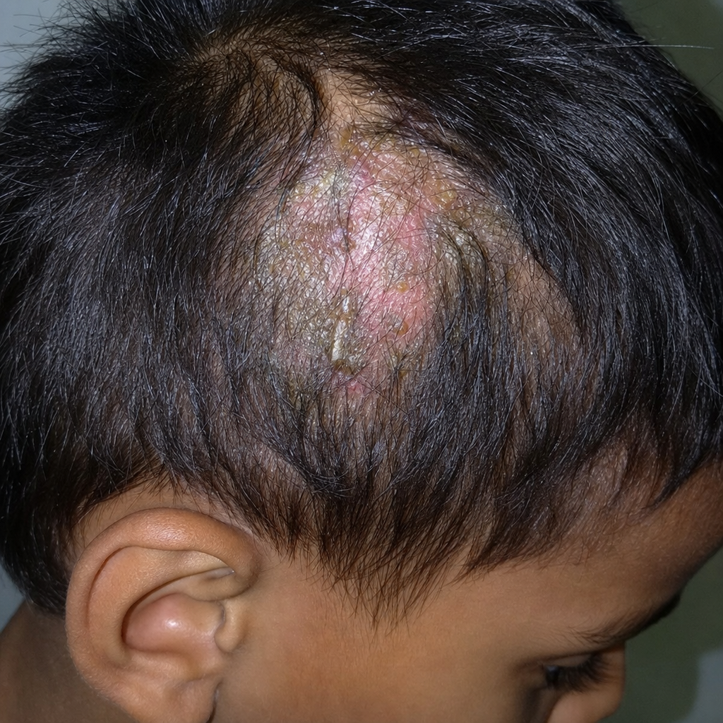
A male child presents with a mildly painful swelling on his scalp for the last 3 months, as shown in the image. The history reveals that there is a pet dog in the household. What is the most likely diagnosis?
A 25-year-old man presents with multiple brownish patches on his trunk that appeared suddenly after exercising in hot weather. The lesions don't itch or scale. Wood's lamp examination shows yellow-green fluorescence. Which of the following is the most appropriate treatment?
All the following drugs are effective in the treatment of Pityriasis Versicolor except:
Selenium sulfide is indicated for treating?
False about Tinea versicolor
Hypersensitivity to dermatophyte antigens is called
A 7-year-old boy presents with patchy hair loss, boggy scalp swelling and broken and fragmented hair follicles at the surface of the scalp resembling black dots. What is the next step in establishing a diagnosis:
An 8-year-old child has localized non-cicatricial alopecia over scalp with itching and scales. The diagnosis is :
Practice by Chapter
Dermatophytoses
Practice Questions
Tinea Versicolor
Practice Questions
Candidiasis
Practice Questions
Onychomycosis
Practice Questions
Subcutaneous Mycoses
Practice Questions
Systemic Mycoses with Cutaneous Manifestations
Practice Questions
Opportunistic Fungal Infections
Practice Questions
Mycetoma
Practice Questions
Tropical Fungal Infections
Practice Questions
Diagnosis of Fungal Infections
Practice Questions
Antifungal Therapy
Practice Questions
Preventive Strategies
Practice Questions
Want unlimited practice?
Get full access to all questions, explanations, and performance tracking.
Scan to download app