
Fungal Skin Infections — MCQs

On this page
A teenage girl presents with multiple white macules on her upper trunk, showing scaling on scraping. KOH mount of the lesions revealed a specific organism. What is the most likely causative agent?
A 16-year-old patient was recently diagnosed with HIV. Which of the following fungal nail infections could be associated?
The appearance of this foot is most consistent with which one of the following diagnoses?
A patient presents with a non-itchy plaque positive for hyphae. What is the most likely diagnosis?
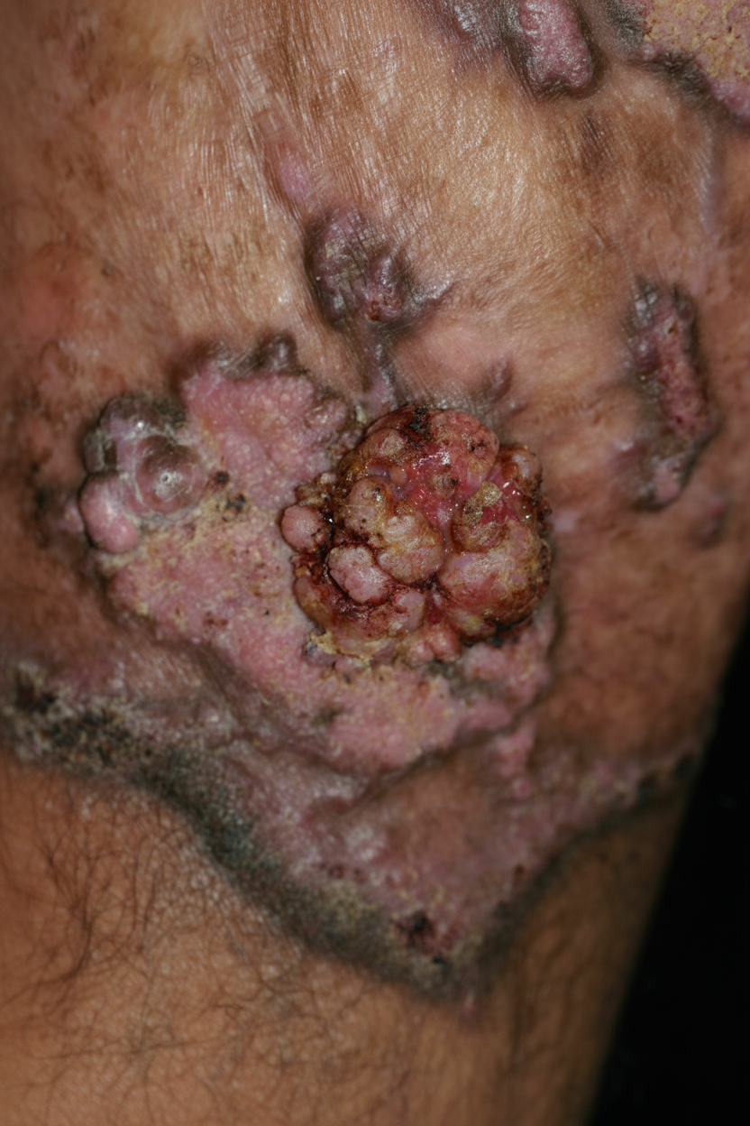
A thirty-two-year-old adult male presents with a chronic suppurative lesion on the angle of the jaw. Microscopic examination shows small abscesses immediately beneath the epidermis with moderate growth of epithelial cords. In the abscess, there are oval, unicellular organisms measuring 20 µm in diameter and having a thick, double-refractive cell wall and cytoplasm containing refractive granules and vacuoles. Which of the following is the most likely diagnosis?
Dermatophytids are defined as:
Which of the following tests is likely to be helpful in diagnosing a patient presenting with an itchy annular plaque on the face?
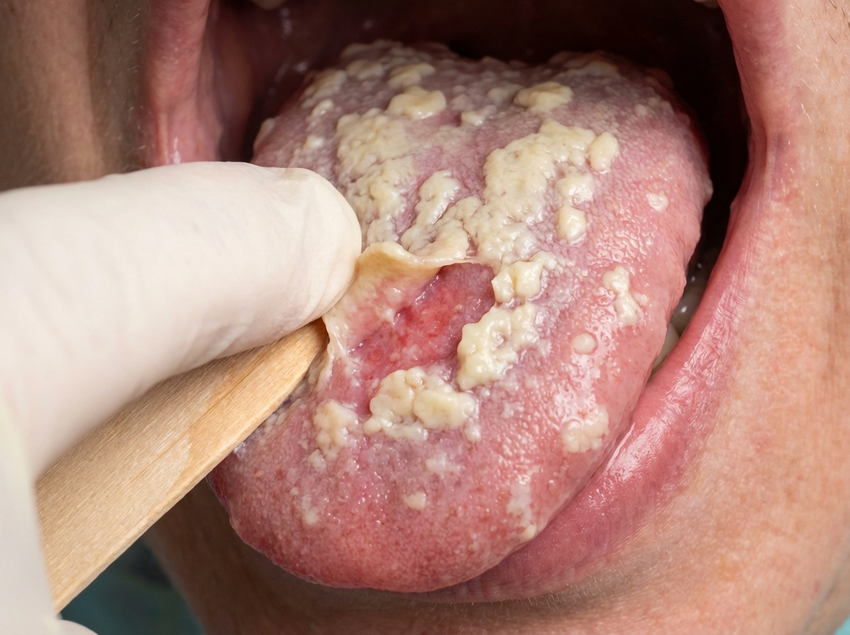
What is the management for the given case?
Potassium iodide is useful in the treatment of which of the following conditions?
In dermatophytosis, which antifungal drug is not indicated?
Practice by Chapter
Dermatophytoses
Practice Questions
Tinea Versicolor
Practice Questions
Candidiasis
Practice Questions
Onychomycosis
Practice Questions
Subcutaneous Mycoses
Practice Questions
Systemic Mycoses with Cutaneous Manifestations
Practice Questions
Opportunistic Fungal Infections
Practice Questions
Mycetoma
Practice Questions
Tropical Fungal Infections
Practice Questions
Diagnosis of Fungal Infections
Practice Questions
Antifungal Therapy
Practice Questions
Preventive Strategies
Practice Questions
Want unlimited practice?
Get full access to all questions, explanations, and performance tracking.
Scan to download app