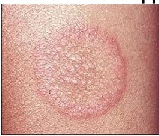
Identify the condition causing this infection on the upper arm

Skin scraping and KOH mounting is primarily used to diagnose which of the following conditions?
Which of the following drugs is effective in the treatment of pityriasis versicolor?
Which of the following fungi is primarily responsible for causing Tinea cruris?
Which of the following is NOT a characteristic of dermatophytosis?
An adult presents with oval, scaly, hypopigmented macules over the chest and back. The diagnosis is
Kerion is a type of:
A 12-year-old girl presents with a well-defined annular pruritic lesion over the neck, with central clearing, that is gradually progressive over the last 8 months. The likely diagnosis is what?
Tinea cruris is caused by which of the following fungi?
Tinea unguium affects
Explanation: ***Tinea corporis (body ringworm)*** - This lesion, depicted on the upper arm, is characteristic of **tinea corporis** due to its **annular, erythematous, and scaly border with central clearing**. - The term "corporis" refers to the **body surface**, excluding the scalp, hands, feet, groin, and nails. *Tinea capitis (scalp ringworm)* - Tinea capitis specifically affects the **scalp** and can present with scaling, hair loss, and inflammation. - The image clearly shows a lesion on the **upper arm**, not the scalp. *Tinea cruris (jock itch)* - Tinea cruris is a fungal infection found in the **groin area**, often extending to the inner thighs and buttocks. - The location of the lesion in the image, on the **upper arm**, rules out tinea cruris. *Tinea manus (hand ringworm)* - Tinea manus affects the **hands**, typically causing dryness, scaling, and sometimes blister formation on the palms or between the fingers. - The lesion in the image is located on the **upper arm**, not the hand.
Explanation: ***Dermatophyte infection*** - Skin scraping followed by **KOH mounting** is the **primary and gold standard** diagnostic method for dermatophyte infections - **Potassium hydroxide (KOH)** dissolves keratin, making **fungal hyphae and arthroconidia** clearly visible under microscopy - This is the **most common indication** for KOH preparation in dermatology practice - Demonstrates **septate hyphae** characteristic of dermatophytes *Candidiasis* - KOH mounting can also identify *Candida* species by revealing **pseudohyphae and budding yeast cells** - While KOH can diagnose candidiasis, dermatophyte infections remain the **primary and most frequent indication** for this test in skin lesions - Candida more commonly affects mucosal surfaces where other diagnostic methods may be preferred *Bacterial infection* - Bacterial infections are diagnosed using **Gram stain, culture, and sensitivity** testing - KOH mounting is specifically for **fungal elements** and does not visualize bacteria effectively - Bacterial skin infections require different diagnostic approaches *Herpes Simplex Virus (HSV)* - HSV infections are diagnosed using **Tzanck smear** (showing multinucleated giant cells), **viral culture, or PCR** - KOH mounting is exclusively for fungal identification and **cannot detect viruses** - Viral cytopathic effects are not visible with KOH preparation
Explanation: ***Ketoconazole*** - **Ketoconazole** is an **azoles antifungal agent** effective against the *Malassezia* species, the causative agent of **pityriasis versicolor**. - It works by inhibiting the synthesis of **ergosterol**, a crucial component of the fungal cell membrane, leading to its disruption and fungal cell death. *Metronidazole* - **Metronidazole** is an **antibiotic** and **amoebicide** primarily used to treat bacterial and parasitic infections. - It has **no antifungal activity** and is thus ineffective against *Malassezia* or other fungal infections. *Griseofulvin* - **Griseofulvin** is an **oral antifungal drug** primarily used for **dermatophyte infections** of the skin, hair, and nails (e.g., tinea capitis, onychomycosis). - It is **ineffective against yeasts and molds**, including *Malassezia*, making it unsuitable for pityriasis versicolor. *Chloroquine* - **Chloroquine** is an **antimalarial drug** also used in the treatment of some autoimmune conditions like lupus erythematosus. - It possesses **no antifungal properties** and is not used to treat fungal infections of any kind.
Explanation: ***Trichophyton rubrum*** - **Trichophyton rubrum** is the **most common dermatophyte** responsible for causing **Tinea cruris**, also known as **jock itch**, accounting for approximately 80-90% of cases. - This fungus thrives in warm, moist environments, making the groin area an ideal site for infection. - It is the **primary causative agent** worldwide for this condition. *Epidermophyton floccosum* - While **Epidermophyton floccosum** can cause **Tinea cruris**, it is significantly less common than **Trichophyton rubrum**, accounting for approximately 10-20% of cases. - It is also known to cause **Tinea pedis** (athlete's foot) and onychomycosis. *Microsporum canis* - **Microsporum canis** is a common cause of **Tinea capitis** (ringworm of the scalp) and **Tinea corporis** (ringworm of the body), particularly in children. - It is primarily transmitted from **infected animals**, especially cats and dogs, to humans and is **rarely implicated in Tinea cruris**. *None of the options* - This option is incorrect because **Trichophyton rubrum** is the well-established and primary cause of **Tinea cruris**. - At least one of the listed options is a recognized pathogen for this condition.
Explanation: ***Subdermal infection*** - Dermatophytosis, or **ringworm**, is characterized by infection of the **superficial keratinized tissues** (skin, hair, nails) and does not typically extend into the subdermal layers. - While fungal infections can be systemic or deep, dermatophytes specifically are restricted to the **stratum corneum** and other dead keratinized structures. *Scaly skin* - **Scaling** is a very common characteristic of dermatophyte infections due to the fungus proliferating within the **stratum corneum**, leading to increased epidermal turnover and shedding. - The scaling can be fine or coarse, often presenting in an **annular (ring-like)** pattern. *Itchy skin* - **Pruritus (itching)** is a prominent symptom of dermatophytosis, often leading patients to seek medical attention. - The itching can range from mild to severe, contributing to discomfort and potential secondary skin excoriations. *Superficial infection* - Dermatophytosis is by definition a **superficial fungal infection**, meaning it is confined to the outermost layers of the skin, hair, and nails. - These fungi produce enzymes such as **keratinases** that allow them to digest keratin, but they generally do not invade viable tissue below the epidermis.
Explanation: ***Pityriasis Versicolor*** - This condition is caused by a **Malassezia species** yeast infection, presenting as characteristic **oval, scaly, hypopigmented or hyperpigmented macules** primarily on the trunk. - The hypopigmentation is often due to the production of **azelaic acid** by the yeast, which inhibits melanin synthesis. *Tuberculosis Cutis* - This refers to various skin manifestations of **tuberculosis**, which can include papules, nodules, ulcers, or verrucous lesions, often with significant inflammation or necrosis. - It does not typically present as widespread, oval, scaly, hypopigmented macules without other systemic signs of tuberculosis. *Psoriasis* - Characterized by **well-demarcated, erythematous plaques** covered with silvery scales, often found on extensor surfaces, scalp, and nails. - **Psoriasis** lesions are typically red and inflamed, not hypopigmented, and do not usually have the fine, scaly appearance described. *Granuloma Annulare* - Presents as **annular (ring-shaped) papules or plaques**, often flesh-colored, red, or pink, most commonly on the hands, feet, and ankles. - It is typically a **non-scaly eruption** and does not cause hypopigmented macules over the trunk.
Explanation: ***Fungal infection*** - Kerion is a severe inflammatory **tinea capitis** (ringworm of the scalp) caused by a fungal infection. - It presents as a boggy, elevated, and painful lesion with pustules and could lead to **scarring alopecia**. *Viral infection* - Viral infections typically manifest with conditions like **herpes zoster**, **varicella**, or **molluscum contagiosum**, which have distinct clinical features and are not characterized as kerion. - Kerion's inflammatory, pustular nature and typical location on the scalp are not consistent with common viral dermatoses. *Bacterial infection* - Bacterial infections of the skin could cause conditions like **impetigo** or **folliculitis**, which are generally less extensive and lack the specific inflammatory boggy plaque typical of kerion. - While secondary bacterial infection can occur in kerion, the primary etiology is fungal, not bacterial. *Parasitic infection* - Parasitic infections of the scalp usually involve **lice** (pediculosis capitis) or **scabies**, which cause intense itching and excoriations but do not result in a kerion-like boggy, inflammatory mass. - The pathology of kerion involves a fungal invasion of hair follicles, distinct from parasitic infestation.
Explanation: ***Tinea corporis*** - This diagnosis is strongly supported by the presentation of a **well-defined, annular, pruritic lesion** with **central clearing**, which is characteristic of dermatophyte infections. - The gradual progression over 8 months is consistent with a chronic fungal infection in an otherwise healthy individual. *Cutaneous tuberculosis* - This condition typically presents with a **nodular, ulcerative, or verrucous lesion**, which is different from the annular lesion with central clearing described. - While it can be chronic, it is often associated with other signs of tuberculosis or immunosuppression, which are not mentioned. *Cutaneous leishmaniasis* - This typically presents as a **papule** enlarging into a **nodule, plaque, or ulcer**, often with a crusted center. - Although it can be chronic, its morphology is generally not described as an annular lesion with central clearing. *Psoriasis* - Psoriasis typically presents with **well-demarcated erythematous plaques** covered by **silvery scales**, usually without central clearing. - While it can be chronic and pruritic, the annular morphology with central clearing is not characteristic of typical psoriasis.
Explanation: ***Epidermophyton*** - **_Epidermophyton floccosum_** is one of the common dermatophytes responsible for causing **tinea cruris** (jock itch) and tinea pedis (athlete's foot). - **Note:** Tinea cruris can be caused by multiple dermatophytes including **_Trichophyton rubrum_** (most common), **_T. mentagrophytes_**, and **_E. floccosum_**. Among the options listed, Epidermophyton is the only dermatophyte that commonly causes tinea cruris. - Microscopic examination of skin scrapings shows **septate hyphae**, and it typically invades the stratum corneum but **not hair or nails**. *Trichosporon* - **_Trichosporon_** species cause **white piedra** (a fungal infection of the hair shaft) and can cause systemic infections in immunocompromised individuals. - It is **not a dermatophyte** and does not cause tinea cruris. *Microsporum* - **_Microsporum_** species are primarily associated with **tinea capitis** (ringworm of the scalp) and **tinea corporis** (ringworm of the body). - While Microsporum can occasionally involve skin in the groin region, it is **rarely implicated in classic tinea cruris** and is not considered a typical causative agent compared to Trichophyton or Epidermophyton species. *Candida* - **_Candida_** species (e.g., **_Candida albicans_**) cause candidiasis, which commonly presents as **intertrigo** in skin folds, oral thrush, or vaginal yeast infections. - While it can occur in the groin area (**candidal intertrigo**), it is **not a dermatophyte** and is distinctly different from tinea cruris, often presenting with **satellite lesions** and an intensely erythematous, macerated rash.
Explanation: ***Nail plate*** - **Tinea unguium**, also known as **onychomycosis**, primarily affects the **nail plate**, causing discoloration, thickening, and crumbling. - The infection starts in the nail bed and invades the nail plate, often leading to **onycholysis** (separation of the nail from its bed). *Nail fold* - Infections of the **nail fold** are typically known as **paronychia**, which is often bacterial or yeast in origin. - While fungal infections can sometimes involve the nail folds, **tinea unguium** specifically refers to fungal infection of the nail plate itself, not primarily the surrounding skin. *Joints* - Fungal infections can rarely cause **septic arthritis**, but this is not what is referred to as **tinea unguium**. - **Tinea unguium** is a superficial fungal infection and does not involve the deeper structures like joints. *Inter digital space* - **Tinea pedis** (athlete's foot) commonly affects the **interdigital spaces** of the feet, causing itching, scaling, and cracking. - While **tinea pedis** can be a risk factor for developing **tinea unguium**, **tinea unguium** itself is specific to the nail and does not primarily manifest in the interdigital spaces.
Dermatophytoses
Practice Questions
Tinea Versicolor
Practice Questions
Candidiasis
Practice Questions
Onychomycosis
Practice Questions
Subcutaneous Mycoses
Practice Questions
Systemic Mycoses with Cutaneous Manifestations
Practice Questions
Opportunistic Fungal Infections
Practice Questions
Mycetoma
Practice Questions
Tropical Fungal Infections
Practice Questions
Diagnosis of Fungal Infections
Practice Questions
Antifungal Therapy
Practice Questions
Preventive Strategies
Practice Questions
Get full access to all questions, explanations, and performance tracking.
Start For Free