
Fungal Skin Infections — MCQs

On this page
Which of the following is NOT a characteristic of dermatophytosis?
Kerion is a type of:
An adult presents with oval, scaly, hypopigmented macules over the chest and back. The diagnosis is
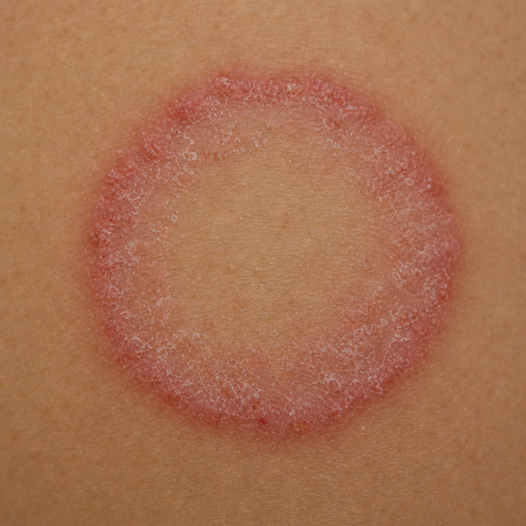
A 12-year-old girl presents with a well-defined annular pruritic lesion over the neck, with central clearing, that is gradually progressive over the last 8 months. The likely diagnosis is what?
What is the treatment for a baby brought to the clinic with a history of hair fall and a boggy scalp with easily pluckable hair, with a similar history occurring six months prior?
Tinea unguium affects
A patient presents with annular, scaly, itchy lesions with central clearing. The most likely causative organism is:
An eleven-year-old boy has Tinea capitis on his scalp. Which of the following is the most appropriate line of treatment for this condition?
What is the correct term for candidiasis of the penis?
In adult patients with multiple scaly macules over the chest and back, which single test is the most comprehensive for diagnosing pityriasis versicolor?
Practice by Chapter
Dermatophytoses
Practice Questions
Tinea Versicolor
Practice Questions
Candidiasis
Practice Questions
Onychomycosis
Practice Questions
Subcutaneous Mycoses
Practice Questions
Systemic Mycoses with Cutaneous Manifestations
Practice Questions
Opportunistic Fungal Infections
Practice Questions
Mycetoma
Practice Questions
Tropical Fungal Infections
Practice Questions
Diagnosis of Fungal Infections
Practice Questions
Antifungal Therapy
Practice Questions
Preventive Strategies
Practice Questions
Want unlimited practice?
Get full access to all questions, explanations, and performance tracking.
Scan to download app