
Fungal Skin Infections — MCQs

On this page
What is the drug of choice for treating onychomycosis?
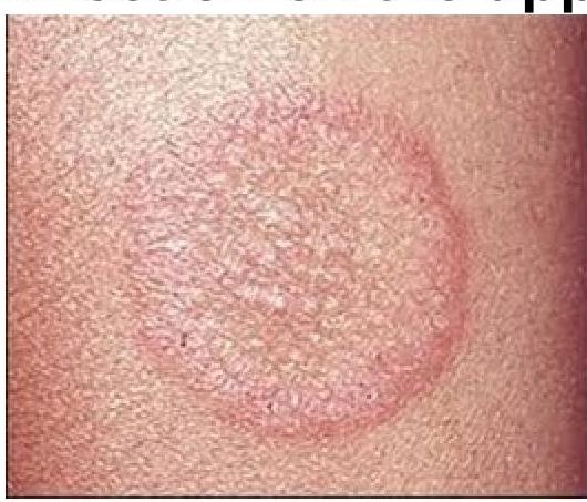
A patient presents with a red, itchy, annular rash with central clearing on the upper arm. Identify the most likely causative organism.
A 40-year-old male patient presents with multiple erythematous annular lesions with peripheral scales arranged predominantly on the trunk. What is the most appropriate treatment option?
What is the primary cause of jock itch?
Which of the following is not a clinical feature of candidal intertrigo?
Most common pattern of onychomycosis is?
Onychomycosis is most commonly caused by which of the following fungi?
Identify the condition causing this infection on the upper arm
Skin scraping and KOH mounting is primarily used to diagnose which of the following conditions?
Which of the following drugs is effective in the treatment of pityriasis versicolor?
Practice by Chapter
Dermatophytoses
Practice Questions
Tinea Versicolor
Practice Questions
Candidiasis
Practice Questions
Onychomycosis
Practice Questions
Subcutaneous Mycoses
Practice Questions
Systemic Mycoses with Cutaneous Manifestations
Practice Questions
Opportunistic Fungal Infections
Practice Questions
Mycetoma
Practice Questions
Tropical Fungal Infections
Practice Questions
Diagnosis of Fungal Infections
Practice Questions
Antifungal Therapy
Practice Questions
Preventive Strategies
Practice Questions
Want unlimited practice?
Get full access to all questions, explanations, and performance tracking.
Scan to download app