Which cellular component is responsible for providing cell shape and motility?
Which force does NOT act in an enzyme-substrate complex?
Trypsinogen is converted to trypsin by which of the following?
What is true about isoenzymes?
Which of the following is a non-competitive inhibitor of intestinal alkaline phosphatase?
What is the specific inhibitor of succinate dehydrogenase?
All of the following could include the mechanism or function of oxygenases, EXCEPT:
Identify the type of inhibition shown in the graph.
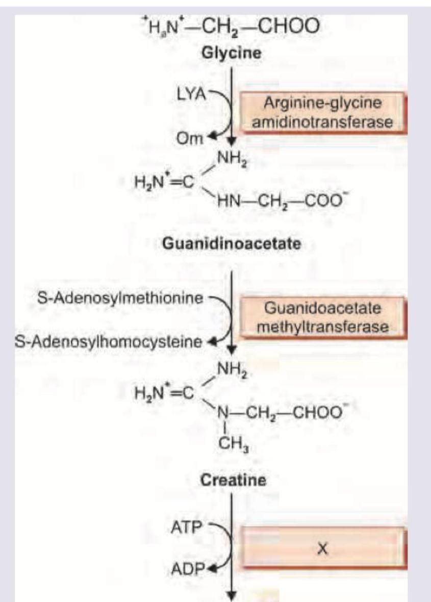
The image shows a biochemical pathway. Name the enzyme marked as X.

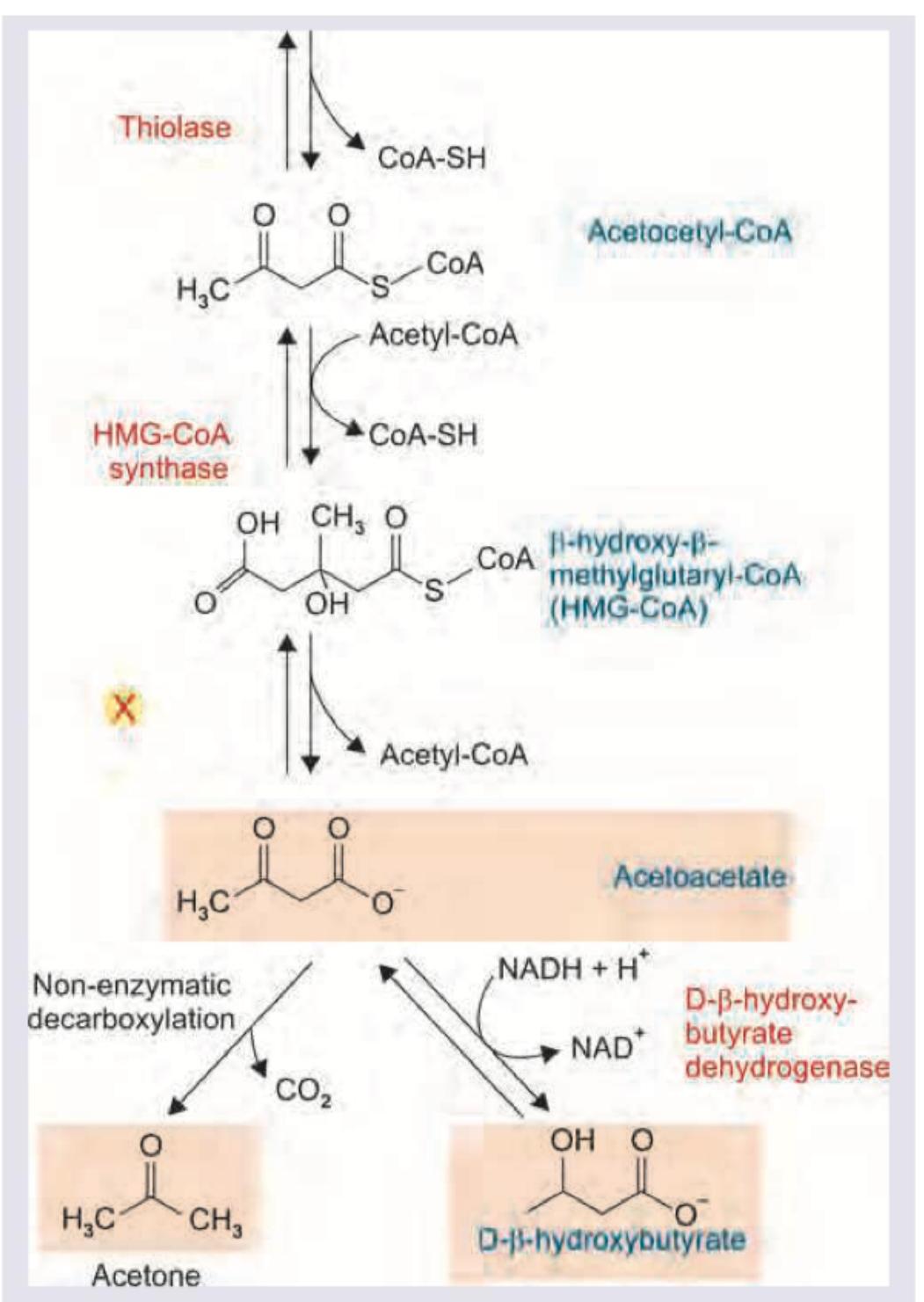
Name the enzyme marked as X in the reaction shown below.

Explanation: **Explanation:** The cytoskeleton is a dynamic network of protein filaments that maintains cellular architecture and facilitates movement. **Microtubules**, composed of $\alpha$ and $\beta$-tubulin dimers, are the thickest components of the cytoskeleton. They are primarily responsible for maintaining **cell shape** (acting as a structural scaffold), enabling **motility** (via cilia and flagella), and facilitating intracellular transport (serving as tracks for dynein and kinesin motors). They also form the mitotic spindle during cell division. **Analysis of Options:** * **Microfilaments (Option A):** Composed of actin, these are primarily involved in muscle contraction, cytokinesis, and maintaining the structure of microvilli. While they contribute to cell shape, microtubules are the primary structural "girders" for overall cell morphology and long-distance motility. * **Golgi Apparatus (Option C):** This organelle is responsible for the post-translational modification, sorting, and packaging of proteins; it does not provide structural shape or motility. * **Mitochondria (Option D):** Known as the "powerhouse of the cell," its primary role is ATP production via oxidative phosphorylation. **High-Yield Clinical Pearls for NEET-PG:** * **Drugs targeting Microtubules:** Remember the mnemonic **"Microtubules Get Vine-Caked"** (Griseofulvin, Vincristine/Vinblastine, Colchicine, Albendazole/Mebendazole, Paclitaxel). * **Kartagener Syndrome:** A triad of situs inversus, chronic sinusitis, and bronchiectasis caused by a defect in **dynein arms** within microtubules, leading to immotile cilia. * **Chediak-Higashi Syndrome:** Involves a defect in microtubule polymerization, leading to impaired phagocytosis and giant lysosomal granules.
Explanation: **Explanation:** The formation of an **Enzyme-Substrate (ES) complex** is primarily driven by weak, non-covalent interactions that allow for rapid binding and release. **1. Why Van der Waals is the Correct Answer:** In the context of standard NEET-PG biochemistry, **Van der Waals forces** are often considered negligible or "non-acting" compared to the stronger, more specific interactions like hydrogen bonding or electrostatic forces. While Van der Waals forces exist between all molecules, they are extremely weak and non-specific. In many classic biochemical models (like the Lock and Key or Induced Fit), the focus is on the specific directional forces that stabilize the transition state. *Note: In advanced biophysics, these forces do exist, but for competitive exams, they are frequently cited as the least significant or "absent" relative to the primary binding forces.* **2. Analysis of Incorrect Options:** * **A. Electrostatic:** These are salt bridges between oppositely charged amino acid side chains (e.g., Aspartate) and the substrate. They are crucial for initial substrate recognition. * **B. Covalent:** While most ES complexes are non-covalent, many enzymes form **transient covalent intermediates** (e.g., Serine proteases like Chymotrypsin). This is a high-yield concept in "Covalent Catalysis." * **D. Hydrogen:** This is the most common force stabilizing the ES complex, providing the specificity required for the enzyme to "recognize" its substrate. **High-Yield Clinical Pearls for NEET-PG:** * **Binding Energy:** The energy released from these weak interactions is called "Binding Energy," which enzymes use to lower the **Activation Energy**. * **Transition State:** Enzymes bind most tightly to the **transition state**, not the substrate itself (Linus Pauling’s principle). * **Irreversible Inhibition:** Drugs like **Aspirin** (inhibiting COX) or **Organophosphates** (inhibiting AChE) work by forming permanent covalent bonds, unlike the transient bonds in normal ES complexes.
Explanation: **Explanation:** **1. Why Enteropeptidase is Correct:** Trypsinogen is an inactive zymogen secreted by the exocrine pancreas. To prevent autodigestion of the pancreas, it must only be activated once it reaches the duodenum. **Enteropeptidase** (also known as enterokinase), an enzyme secreted by the duodenal mucosal cells (Brunner’s glands), acts as the specific "molecular switch." It cleaves a hexapeptide from the N-terminal end of trypsinogen, converting it into its active form, **trypsin**. Once a small amount of trypsin is formed, it acts autocatalytically to activate more trypsinogen and other pancreatic zymogens (chymotrypsinogen, procarboxypeptidase, and proelastase). **2. Why Other Options are Incorrect:** * **B. Acidic pH:** Acidic pH (gastric acid) is responsible for activating **pepsinogen to pepsin** in the stomach. In contrast, pancreatic enzymes like trypsin require an alkaline pH (provided by bicarbonate) to function optimally. * **C. Elastase:** Elastase is a protease itself, but it is secreted as the zymogen **proelastase**, which requires active trypsin for its activation. It does not activate trypsinogen. **3. Clinical Pearls & High-Yield Facts for NEET-PG:** * **The "Master Switch":** Trypsin is considered the master activator of all pancreatic proteases. * **Deficiency:** Congenital enteropeptidase deficiency leads to severe protein malabsorption and failure to thrive because no pancreatic proteases can be activated. * **Protection Mechanism:** The pancreas also secretes **PSTI (Pancreatic Secretory Trypsin Inhibitor)** to neutralize any trypsin prematurely activated within the pancreatic ducts, preventing acute pancreatitis. * **Localization:** Enteropeptidase is located on the brush border of the duodenal enterocytes.
Explanation: **Explanation:** Isoenzymes (or isozymes) are physically distinct forms of the same enzyme that catalyze the same chemical reaction but differ in their amino acid sequences and biochemical properties. **Why Option D is Correct:** * **Different Km values (Option A):** Isoenzymes often have different affinities for the same substrate. A classic example is **Glucokinase (Hexokinase IV)** and **Hexokinase I**. Glucokinase has a high Km (low affinity) for glucose, allowing it to function only when blood glucose levels are high, whereas Hexokinase I has a low Km (high affinity) to ensure glucose uptake even during fasting. * **Multimeric complexes (Option B):** Many isoenzymes are oligomeric, formed by different combinations of polypeptide subunits. For instance, **LDH (Lactate Dehydrogenase)** is a tetramer composed of H (Heart) and M (Muscle) subunits, resulting in five distinct isoenzymes (LDH1 to LDH5). **CK (Creatine Kinase)** is a dimer (B and M subunits) forming CK-BB, CK-MB, and CK-MM. * **Different physical properties (Option C):** Due to variations in amino acid composition, isoenzymes exhibit different electrophoretic mobilities, heat stabilities, and response to inhibitors. This allows them to be separated and quantified in a clinical laboratory. **Clinical Pearls for NEET-PG:** 1. **LDH Flip:** Normally LDH1 < LDH2. In **Myocardial Infarction (MI)**, LDH1 becomes greater than LDH2 (LDH Flip). 2. **CK-MB:** This is the specific marker for MI; it rises within 4-8 hours and returns to baseline within 48-72 hours. 3. **Alkaline Phosphatase (ALP):** Isoenzymes include Regan (carcinoplacental), heat-stable (placental), and bone-specific forms. 4. **Tissue Specificity:** Isoenzymes allow for fine-tuning of metabolism to meet the specific physiological needs of different organs.
Explanation: **Explanation:** Alkaline Phosphatase (ALP) exists in several tissue-specific isoenzymes (liver, bone, kidney, placenta, and intestine). A unique biochemical characteristic of these isoenzymes is their susceptibility to specific amino acid inhibitors, which is often used in laboratory medicine to differentiate the source of elevated ALP levels. **1. Why L-Phenylalanine is correct:** L-Phenylalanine is a classic **uncompetitive/non-competitive inhibitor** specifically for the **intestinal** and **placental** isoenzymes of ALP. It binds to the enzyme-substrate complex, preventing the reaction from proceeding. This inhibition is highly specific; it does not affect the liver, bone, or kidney (tissue non-specific) isoenzymes. In clinical biochemistry, adding L-phenylalanine to a serum sample helps quantify the proportion of intestinal ALP present. **2. Why other options are incorrect:** * **L-Alanine:** This amino acid is a specific inhibitor of the **liver and kidney** isoenzymes of ALP, rather than the intestinal form. * **L-Tyrosine & L-Tryptophan:** While these are aromatic amino acids like phenylalanine, they do not exhibit the same potent or specific inhibitory effect on intestinal ALP isoenzymes and are not used as diagnostic markers in this context. **High-Yield Clinical Pearls for NEET-PG:** * **Heat Stability Test:** This is another method to differentiate ALP. **Placental ALP** is the most heat-stable ("Regan Isoenzyme"), while **Bone ALP** is the most heat-labile ("Bone burns"). * **Levamisole:** Inhibits all ALP isoenzymes *except* intestinal and placental forms. * **Clinical Significance:** Intestinal ALP is often elevated in individuals with blood groups B or O after a fatty meal and in patients with cirrhosis.
Explanation: **Explanation:** **1. Why Malonate is the Correct Answer:** Malonate is the classic example of a **competitive inhibitor**. It is a structural analogue of **succinate**, the substrate for **Succinate Dehydrogenase (SDH)** in the Citric Acid Cycle (TCA). Because malonate mimics the shape of succinate, it competes for the same active site on the enzyme. This inhibition can be overcome by increasing the concentration of the substrate (succinate). SDH is unique because it is the only TCA cycle enzyme embedded in the inner mitochondrial membrane, also functioning as **Complex II** of the Electron Transport Chain (ETC). **2. Why the Other Options are Incorrect:** * **A. Cyanide:** This is a potent inhibitor of **Complex IV (Cytochrome c oxidase)** in the ETC. It binds to the ferric iron ($Fe^{3+}$) in the heme group, halting cellular respiration. * **C. Arsenite:** This inhibits enzymes that require **Lipoic acid** as a cofactor, most notably the **Pyruvate Dehydrogenase (PDH) complex** and $\alpha$-ketoglutarate dehydrogenase. It binds to the -SH groups of lipoic acid. * **D. Fluoride:** This is a specific inhibitor of **Enolase** in the Glycolysis pathway. In clinical practice, fluoride is added to blood collection tubes (grey top) to prevent glycolysis before glucose estimation. **3. High-Yield Clinical Pearls for NEET-PG:** * **SDH Marker:** Succinate dehydrogenase is a marker enzyme for the **mitochondria**. * **Competitive Inhibition:** In the presence of malonate, the **$K_m$ increases** (affinity decreases) while the **$V_{max}$ remains unchanged**. * **Complex II:** Unlike Complexes I, III, and IV, Complex II (SDH) does not pump protons across the mitochondrial membrane.
Explanation: **Explanation:** **Oxygenases** are a class of enzymes that catalyze the direct incorporation of oxygen into a substrate molecule. **Why Option D is the Correct Answer (The Exception):** Oxygenases are involved in **hydroxylation** and **oxygenation** reactions, not carboxylation. **Carboxylation** (the addition of $CO_2$) is catalyzed by **Carboxylases**, which typically require **Biotin** as a cofactor and ATP as an energy source (e.g., Pyruvate carboxylase). The metabolism of drugs in the liver (Phase I reactions) primarily involves hydroxylation via the Cytochrome P450 system, which is a type of oxygenase, not a carboxylase. **Analysis of Incorrect Options:** * **Option A (Incorporate 2 atoms of oxygen):** This describes **Dioxygenases** (e.g., Tryptophan pyrrolase, Homogentisate oxidase). They incorporate both atoms of an $O_2$ molecule into the substrate ($S + O_2 \rightarrow SO_2$). * **Option B (Incorporate 1 atom of oxygen):** This describes **Monooxygenases** (also known as Mixed-Function Oxidases). They incorporate one atom of oxygen into the substrate as a hydroxyl group and reduce the other atom to water ($S + O_2 + ZH_2 \rightarrow S-OH + H_2O + Z$). * **Option C (Required for hydroxylation of steroids):** This is a classic function of monooxygenases. The **Cytochrome P450** system in the adrenal cortex and gonads is essential for the hydroxylation steps in steroid hormone synthesis. **High-Yield Clinical Pearls for NEET-PG:** * **Cytochrome P450 (CYP450):** The most significant monooxygenase system, located in the endoplasmic reticulum (microsomes). It is vital for drug detoxification and steroidogenesis. * **Phenylalanine Hydroxylase:** A clinically important monooxygenase; its deficiency leads to **Phenylketonuria (PKU)**. * **Key Cofactors:** Monooxygenases often require **NADPH**, **FAD**, or **Tetrahydrobiopterin ($BH_4$)** as electron donors.
Explanation: ***Noncompetitive inhibition***- This inhibition type is characterized by a decrease in **$V_{max}$** but no change in the **$K_m$** value, meaning the inhibitor reduces the enzyme's efficiency but not its affinity for the substrate.- The inhibitor typically binds reversibly to an **allosteric site** (not the active site), affecting the enzyme's catalytic functionality whether the substrate is bound or not.*Competitive inhibition*- Competitive inhibition is characterized by an **increased $K_m$** (decreased apparent affinity) while the **$V_{max}$** remains unchanged.- The inhibitor binds directly to the **active site**, competing with the substrate, and the effect can be overcome by increasing substrate concentration.*Allosteric Inhibition*- Allosteric inhibition is a general mechanism where a molecule binds to a site other than the active site (**allosteric site**), changing the enzyme's conformation and activity.- While noncompetitive and uncompetitive inhibitions are types of allosteric regulation, "noncompetitive inhibition" is the specific and most accurate term for the observed kinetic behavior (decreased $V_{max}$, constant $K_m$).*Uncompetitive inhibition*- This type involves the inhibitor binding only to the **enzyme-substrate complex (ES)**, resulting in a proportional decrease in both **$V_{max}$** and **$K_m$**.- On a Lineweaver-Burk plot, this is shown by parallel lines, highly distinguishing it from noncompetitive inhibition where the lines intersect on the X-axis.
Explanation: ***Creatine Kinase*** - The image shows the synthesis of creatine, and the final step involves the conversion of **creatine** to **creatine phosphate** using **ATP**, which is typical of a kinase enzyme. - **Creatine kinase** (CK) catalyzes the reversible transfer of a phosphate group from ATP to creatine, forming phosphocreatine and ADP, or vice versa, playing a crucial role in energy storage in muscle. *Creatine Phosphorylase* - This term is not a standard or commonly recognized name for an enzyme in biochemistry. While it might sound related to phosphorylation of creatine, **creatine kinase (CK)** is the correct and widely accepted enzyme name. - Enzymes are named based on their function and substrate; "phosphorylase" usually refers to enzymes that cleave bonds using inorganic phosphate, which is not the reaction shown here. *Argininosuccinase* - **Argininosuccinase (argininosuccinate lyase)** is an enzyme involved in the **urea cycle**, catalyzing the cleavage of argininosuccinate into arginine and fumarate. - This enzyme is not involved in creatine metabolism and its reaction mechanism is distinct from the phosphorylation shown in the diagram. *Ornithine transcarbamoylase* - **Ornithine transcarbamoylase** (OTC) is another enzyme of the **urea cycle**, catalyzing the reaction between carbamoyl phosphate and ornithine to form citrulline. - Its function is entirely unrelated to the synthesis or phosphorylation of creatine and does not involve ATP-ADP interconversion in this manner.
Explanation: ***HMG CoA Lyase*** - The reaction shown converts **HMG-CoA** into **acetoacetate** and **acetyl-CoA**. This cleavage reaction is catalyzed by **HMG-CoA lyase**. - This enzyme is crucial in **ketogenesis**, the metabolic pathway that produces ketone bodies. *HMG COA reductase* - **HMG-CoA reductase** is involved in the synthesis of **cholesterol**, not the breakdown of HMG-CoA into acetoacetate. - It catalyzes the reduction of HMG-CoA to **mevalonate**, which is a precursor for cholesterol and isoprenoid synthesis. *HMG COA oxidase* - This enzyme is not a recognized enzyme in the metabolism of HMG-CoA. - The name suggests an oxidation reaction, which is not what occurs when HMG-CoA is converted to acetoacetate. *HMG COA Trans-peptidase* - This enzyme name does not correspond to any known enzyme involved in HMG-CoA metabolism. - Transpeptidases are typically involved in peptide bond formation or rearrangement, which is unrelated to ketone body synthesis.
Enzyme Classification and Nomenclature
Practice Questions
Enzyme Kinetics and Michaelis-Menten Equation
Practice Questions
Enzyme Inhibition: Competitive and Non-competitive
Practice Questions
Allosteric Regulation
Practice Questions
Coenzymes and Cofactors
Practice Questions
Isoenzymes and Clinical Significance
Practice Questions
Enzyme Regulation: Covalent Modification
Practice Questions
Enzyme Regulation: Zymogen Activation
Practice Questions
Enzyme Induction and Repression
Practice Questions
Ribozymes and Catalytic RNA
Practice Questions
Enzyme Diagnostic Applications
Practice Questions
Enzyme Therapy and Inhibitors as Drugs
Practice Questions
Get full access to all questions, explanations, and performance tracking.
Start For Free