
Systemic Anatomy — MCQs

On this page
Which of the following is an example of a syndesmosis joint?
Which of the following is NOT a digastric muscle?
Which of the following muscles is a convergent muscle?
What is the solid and largest lymphatic organ of the body?
Which of the following is a fibrous joint?
Which of the following is the type of joint between epiphysis and diaphysis of a long bone?
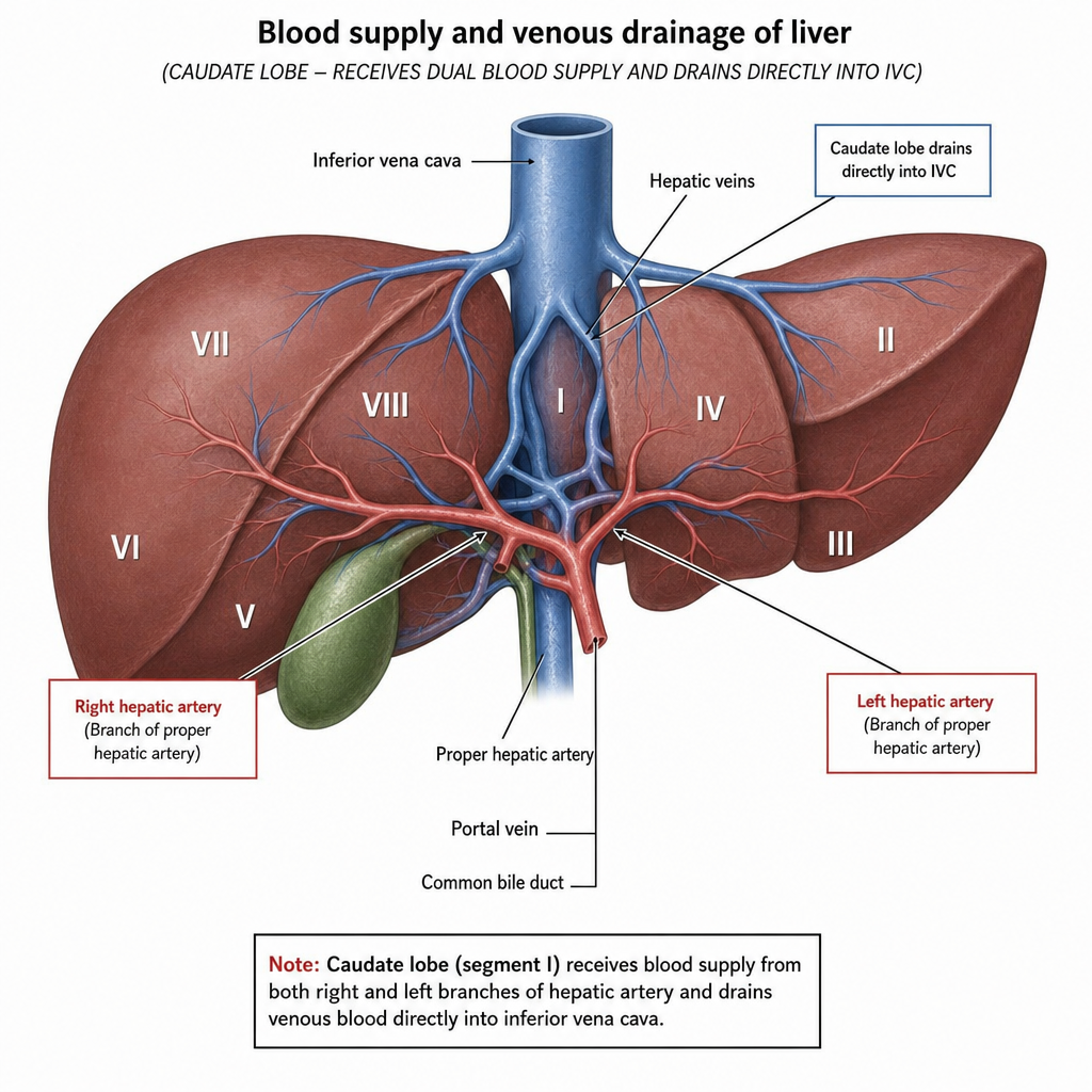
Which of the following segment of liver receives blood from right and left branches of hepatic artery and drains directly into IVC?
Articular surfaces ≥3 are present in which type of joint?
All of the following are composite muscles, except:
Absence of lymph nodes is characteristic of -
Practice by Chapter
Skeletal System
Practice Questions
Articular System
Practice Questions
Muscular System
Practice Questions
Cardiovascular System
Practice Questions
Lymphatic System
Practice Questions
Nervous System
Practice Questions
Respiratory System
Practice Questions
Digestive System
Practice Questions
Urinary System
Practice Questions
Reproductive System
Practice Questions
Endocrine System
Practice Questions
Integumentary System
Practice Questions
Want unlimited practice?
Get full access to all questions, explanations, and performance tracking.
Scan to download app