What is the blood supply of the great toe?
Which drug is contraindicated for use along with local anaesthetics?
Which of the following types of leukemia almost never develops after radiation exposure?
What is the most common nerve involved in cavernous sinus thrombosis?
A fixed firm belief that the patient has subjective certainty is called as?
Which of the following is an example of a traction epiphysis?
Which of the following structures is most likely to be compressed by an aneurysm of the posterior communicating artery?
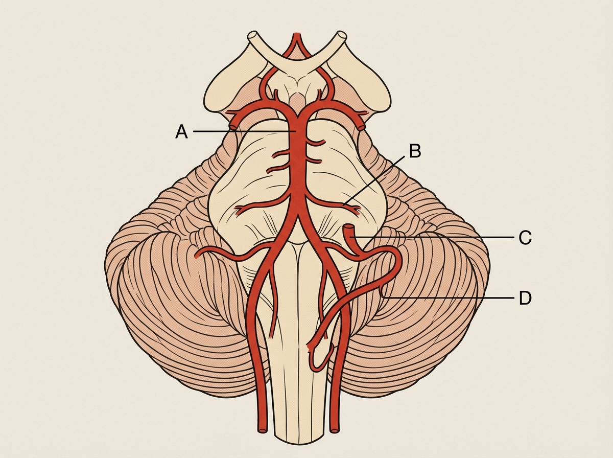
Which artery is involved in Wallenberg syndrome?

Which of the following is a hallmark of acute inflammation?
What is a ganglion?
Explanation: The blood supply to the foot is divided into dorsal and plantar systems. The **Dorsalis pedis artery (DPA)**, a continuation of the anterior tibial artery, is the primary source of blood for the dorsal aspect of the foot and the toes. **Why Dorsalis Pedis Artery is Correct:** Upon reaching the first intermetatarsal space, the DPA gives off the **First Dorsal Metatarsal Artery**. This specific branch further divides to supply the medial and lateral sides of the **great toe (hallux)** and the medial side of the second toe. This makes the DPA the direct arterial source for the great toe's dorsal surface. **Analysis of Incorrect Options:** * **Lateral plantar artery:** This is a branch of the posterior tibial artery. While it forms the deep plantar arch, it primarily supplies the lateral side of the sole and the lateral four toes. * **Metatarsal artery:** While dorsal metatarsal arteries supply the toes, the question asks for the primary parent source. The first metatarsal artery is a direct branch of the DPA, making DPA the most definitive answer. * **Posterior tibial artery:** This artery supplies the sole of the foot via the medial and lateral plantar arteries. While it contributes to the plantar supply of the toes, the DPA is the classic anatomical answer for the primary supply of the hallux. **NEET-PG High-Yield Pearls:** * **Palpation Point:** The DPA pulse is felt on the dorsum of the foot, lateral to the tendon of **Extensor Hallucis Longus (EHL)**. * **Termination:** The DPA terminates by dipping into the first interosseous space to join the lateral plantar artery, completing the **plantar arch**. * **Clinical Significance:** Absence of the DPA pulse is a classic sign of **Peripheral Arterial Disease (PAD)** or Buerger's disease.
Explanation: ### Explanation **Core Concept: Vasoconstriction and Tissue Necrosis** Local anesthetics (LAs) are often combined with vasoconstrictors (like Adrenaline) to prolong the duration of action, decrease systemic toxicity, and provide a bloodless surgical field. However, **Noradrenaline** is contraindicated for this purpose. **Why Noradrenaline is the Correct Answer:** Noradrenaline is a potent **alpha-1 adrenergic agonist** with minimal beta-2 activity. When injected locally, it causes intense, prolonged vasoconstriction. This can lead to severe localized ischemia, tissue hypoxia, and subsequent **tissue necrosis or sloughing** at the injection site. Unlike adrenaline, which has balanced alpha and beta effects (causing some vasodilation in skeletal muscle), noradrenaline’s vasoconstriction is too aggressive for safe local infiltration. **Analysis of Incorrect Options:** * **Adrenaline (Option C):** This is the **gold standard** vasoconstrictor used with LAs (usually in a 1:200,000 concentration). It effectively delays absorption without the extreme ischemic risk associated with noradrenaline. * **Dopamine & Dobutamine (Options A & B):** These are primarily used as inotropic/vasopressor agents in critical care (e.g., shock or heart failure). They are not used as adjuncts to local anesthesia, but they are not specifically "contraindicated" in the same pharmacological context as noradrenaline regarding local tissue necrosis. **High-Yield Clinical Pearls for NEET-PG:** * **The "End-Artery" Rule:** Even with Adrenaline, avoid use in areas supplied by end-arteries (fingers, toes, nose, ears, and penis) to prevent gangrene. * **Maximum Dose:** The addition of Adrenaline increases the maximum safe dose of Lignocaine from **3 mg/kg to 7 mg/kg**. * **Felypressin:** A synthetic vasopressin derivative used as an alternative to adrenaline in dental anesthesia, especially for patients where catecholamines are risky (e.g., hyperthyroidism).
Explanation: The relationship between ionizing radiation and leukemogenesis is well-established; however, not all hematological malignancies share this association. **Why Chronic Lymphocytic Leukemia (CLL) is the correct answer:** CLL is the only major type of leukemia that has **no proven association with radiation exposure**. Large-scale epidemiological studies (including those of Hiroshima and Nagasaki survivors and patients receiving radiotherapy) have consistently shown that the incidence of CLL does not increase following radiation. The etiology of CLL is more closely linked to genetic factors, family history, and advanced age rather than environmental triggers like ionizing radiation or chemicals. **Analysis of Incorrect Options:** * **Acute Myeloblastic Leukemia (AML):** This is the most common radiation-induced leukemia in adults. It typically presents with a peak incidence 5–10 years after exposure. * **Chronic Myeloid Leukemia (CML):** CML shows a strong correlation with radiation. It was one of the first malignancies linked to the survivors of atomic bombings. * **Acute Lymphoblastic Leukemia (ALL):** ALL is the most common radiation-induced leukemia in children. **High-Yield Pearls for NEET-PG:** * **Radiation Sensitivity:** The bone marrow is one of the most radiosensitive tissues in the body. * **Latency Period:** Leukemia has a shorter latency period (2–10 years) compared to solid tumors (10–30 years) following radiation exposure. * **CLL Exception:** Always remember: **"CLL = Constant Low Likelihood"** of being caused by radiation. * **Benzene Exposure:** While radiation doesn't cause CLL, chemical exposure like Benzene is strongly linked to **AML**.
Explanation: **Explanation:** The **Abducens nerve (CN VI)** is the most common and earliest nerve involved in cavernous sinus thrombosis (CST). This is due to its unique anatomical position: while the Oculomotor (CN III), Trochlear (CN IV), and Ophthalmic (V1) nerves are protected within the lateral wall of the cavernous sinus, the Abducens nerve runs **medially through the center** of the sinus, in close proximity to the internal carotid artery. Being "free-floating" within the venous blood of the sinus makes it highly susceptible to compression or inflammation from a thrombus. **Analysis of Options:** * **Abducens nerve (Correct):** Its central location makes it the first to be affected, typically presenting as a loss of lateral gaze (lateral rectus palsy). * **Trochlear nerve (Incorrect):** Located in the lateral wall of the sinus; it is usually involved later as the thrombosis expands. * **Oculomotor nerve (Incorrect):** Also located in the lateral wall; while frequently involved in advanced CST, it is rarely the initial or most common isolated finding. * **Facial nerve (Incorrect):** The Facial nerve (CN VII) does not pass through the cavernous sinus; it exits the skull via the stylomastoid foramen. **High-Yield Clinical Pearls for NEET-PG:** * **Earliest Sign:** The earliest clinical sign of CST is often a **paralysis of the lateral rectus muscle** (CN VI palsy). * **Danger Triangle:** Infections of the "danger area of the face" (nasolabial fold to bridge of nose) can lead to CST via the **superior ophthalmic vein**, which lacks valves. * **Structures in the Lateral Wall (Superior to Inferior):** CN III, CN IV, V1 (Ophthalmic), and V2 (Maxillary). * **Structures passing through the Center:** CN VI and the Internal Carotid Artery.
Explanation: **Explanation:** The correct answer is **Delusion**. In psychiatry and neuroanatomy, a delusion is defined as a **fixed, false belief** that is firmly held despite incontrovertible evidence to the contrary and is not consistent with the patient’s educational, social, or cultural background. The hallmark of a delusion is the "subjective certainty" with which the patient holds the belief, making it impervious to logic or reasoning. **Analysis of Options:** * **Hallucination:** These are sensory perceptions in the absence of an external stimulus (e.g., hearing voices when no one is speaking). While the patient may believe they are real, hallucinations are disorders of *perception*, whereas delusions are disorders of *thought content*. * **Depersonalization:** This is a dissociative symptom where the patient feels detached from themselves, as if they are an outside observer of their own body or mental processes. It is a disorder of *self-awareness*. * **Derealization:** This involves a feeling that the external world is unreal, strange, or "dream-like." Like depersonalization, it is a dissociative phenomenon rather than a fixed belief. **High-Yield Clinical Pearls for NEET-PG:** * **Primary Delusion (Autochthonous):** A delusion that arises suddenly ("out of the blue") without a preceding event. * **Overvalued Idea:** A belief that is held with strong conviction but is not as fixed or irrational as a delusion (often seen in OCD or Anorexia). * **Anatomical Correlation:** Delusional thinking is often associated with dysfunction in the **prefrontal cortex** and imbalances in **dopaminergic pathways** (specifically the mesolimbic pathway). * **Key Distinction:** Delusions = Disorder of **Thought Content**; Loosening of Associations = Disorder of **Thought Form**.
Explanation: An **epiphysis** is the end of a long bone that ossifies from a secondary center [1]. To answer this question, one must understand the classification of epiphyses based on their functional nature: 1. **Traction Epiphysis (Correct Answer: A):** These are non-articular and do not contribute to the longitudinal growth of the bone. They are formed due to the **pull (traction) of powerful muscles** or tendons. The **Greater and Lesser Trochanters** of the femur (gluteal and iliopsoas pull) and the **Tuberosities** of the humerus are classic examples. 2. **Atavistic Epiphysis (Option D):** These represent bones that were phylogenetically independent in lower animals but have fused with other bones in humans. The **Coracoid process** of the scapula and the posterior tubercle of the talus are examples. 3. **Pressure Epiphysis (Option C):** These are articular and transmit the weight of the body. They are found at the ends of long bones [2] (e.g., Head of the femur, Lower end of the radius). **Carpals** and tarsals are short bones that primarily undergo pressure-based ossification. 4. **Aberrant Epiphysis (Option B):** These are epiphyses not always present, such as the epiphysis at the base of the second metacarpal or the **Os trigonum** (which is often an accessory ossicle/unfused atavistic epiphysis). **High-Yield NEET-PG Pearls:** * **Pressure Epiphysis:** Protects the growth plate from compression (e.g., Head of Femur). * **Traction Epiphysis:** Always associated with muscle attachments (e.g., Trochanters, Tuberosities, Epicondyles of humerus). * **Atavistic Epiphysis:** "Evolutionary leftovers" (e.g., Coracoid process, Os trigonum).
Explanation: **Explanation:** The **Oculomotor nerve (CN III)** is the most common cranial nerve affected by intracranial aneurysms, specifically those arising at the junction of the **Posterior Communicating Artery (PCoA)** and the Internal Carotid Artery [2]. **1. Why the Oculomotor Nerve is Correct:** Anatomically, the oculomotor nerve emerges from the midbrain and passes forward in the subarachnoid space, running **parallel and just lateral** to the posterior communicating artery. Because of this extreme proximity, an aneurysm (dilation) of the PCoA directly compresses the nerve. Since the parasympathetic pupilloconstrictor fibers are located superficially (peripherally) in the nerve trunk, they are affected first, leading to a **dilated, non-reactive pupil** before motor paralysis (ptosis and "down and out" eye) occurs [1]. **2. Why the Other Options are Incorrect:** * **Optic Nerve (CN II):** Located more medially and anteriorly; it is more likely to be compressed by an aneurysm of the **Anterior Communicating Artery** or the Ophthalmic artery [2]. * **Trochlear Nerve (CN IV):** Although it also passes between the posterior cerebral and superior cerebellar arteries, it runs more laterally and is significantly further from the PCoA junction than CN III. * **Hypophysis Cerebri (Pituitary Gland):** Located in the sella turcica. While it can be compressed by a large Internal Carotid Artery aneurysm within the cavernous sinus, it is not the primary structure related to the PCoA. **Clinical Pearls for NEET-PG:** * **Medical vs. Surgical Third Nerve Palsy:** PCoA aneurysm causes "Surgical" palsy (pupil involved). Diabetes mellitus causes "Medical" palsy (pupil spared due to microvascular ischemia affecting the central motor fibers). * **Rule of the Pupil:** Any CN III palsy with a dilated pupil is a neurosurgical emergency until a PCoA aneurysm is ruled out. * **Location:** The PCoA connects the Internal Carotid Artery to the Posterior Cerebral Artery in the Circle of Willis [2].
Explanation: ***Artery D*** - **Posterior Inferior Cerebellar Artery (PICA)** occlusion causes **Wallenberg syndrome** (lateral medullary syndrome). - Results in characteristic symptoms including **ipsilateral facial numbness**, **contralateral body numbness**, **dysphagia**, and **ataxia**. *Artery A* - Does not supply the **lateral medulla** where Wallenberg syndrome pathology occurs. - Occlusion would cause different neurological deficits not consistent with **lateral medullary syndrome**. *Artery B* - Not the primary vessel responsible for **lateral medullary** blood supply. - Involvement would produce alternative stroke syndromes without the classic **PICA territory** features. *Artery C* - Does not correspond to the **PICA distribution** necessary for Wallenberg syndrome. - Occlusion would result in different anatomical areas affected, lacking the **dorsolateral medullary** involvement.
Explanation: **Explanation:** Acute inflammation is the immediate and early response to an injurious agent, characterized by three main components: alterations in vascular caliber, structural changes in microvasculature, and emigration of leukocytes. **Why Option C is Correct:** The hallmarks of acute inflammation are **vasodilation** and **increased vascular permeability** [2]. 1. **Vasodilation:** Induced by mediators like histamine and nitric oxide, it leads to increased blood flow (causing heat and redness) [1], [2]. 2. **Increased Permeability:** This is the most characteristic feature. It allows protein-rich fluid (exudate) to move into extravascular tissues, resulting in edema [2], [3]. This is primarily achieved through endothelial cell contraction, creating "gaps" in the post-capillary venules. **Analysis of Incorrect Options:** * **A. Vasoconstriction:** While transient vasoconstriction of arterioles occurs for a few seconds immediately after injury, it is a minor, fleeting event and not a hallmark of the inflammatory process [4]. * **B. Stasis:** Stasis (slowing of blood flow) occurs *as a result* of increased permeability and fluid loss, which increases blood viscosity. It is a secondary phenomenon, not the primary hallmark. * **D. Leukocyte Margination:** This is a cellular event where WBCs move toward the periphery of the vessel wall. While essential for emigration, it is a consequence of stasis and not the defining vascular hallmark. **High-Yield Clinical Pearls for NEET-PG:** * **Cardinal Signs of Inflammation:** Rubor (redness), Calor (heat), Tumor (swelling), Dolor (pain), and Functio Laesa (loss of function) [1]. * **Triple Response of Lewis:** Flush (capillary dilation), Flare (arteriolar dilation), and Wheal (exudation/edema). * **Most common mechanism of vascular leakage:** Endothelial cell contraction (immediate transient response).
Explanation: ### Explanation **1. Why Option B is Correct:** In neuroanatomy, a **ganglion** is defined as a collection of neuronal cell bodies located in the **Peripheral Nervous System (PNS)**—that is, outside the brain and spinal cord [4]. These structures serve as relay stations or processing centers for nerve impulses. Examples include the Dorsal Root Ganglia (sensory) and Sympathetic Chain Ganglia (autonomic) [2], [4]. **2. Why the Other Options are Incorrect:** * **Option A:** A collection of neuronal cell bodies *within* the Central Nervous System (CNS) is called a **Nucleus**. (Exception: The "Basal Ganglia" are located in the CNS [4], but this is a historical misnomer; they are technically nuclei). * **Option C:** A collection of dendrites does not define a ganglion. Ganglia are characterized by the presence of the **soma (cell body)**, which contains the nucleus and Nissl substance [1]. * **Option D:** This is too broad. Neuroanatomy maintains a strict terminological distinction based on location (CNS vs. PNS) to differentiate between nuclei and ganglia. **3. NEET-PG High-Yield Clinical Pearls:** * **Pseudounipolar Neurons:** These are characteristically found in the **Dorsal Root Ganglia (DRG)** and the sensory ganglia of cranial nerves [3]. * **Satellite Cells:** These are the specific glial cells that surround and support neurons within a ganglion. * **Basal Ganglia Exception:** Always remember for exams that the Basal Ganglia are the only major "ganglia" located inside the CNS (involved in motor control) [4]. * **Autonomic Ganglia:** These are sites of synapse between preganglionic and postganglionic fibers (e.g., Ciliary, Pterygopalatine, Submandibular, and Otic ganglia in the head and neck) [4].
Organization of the Nervous System
Practice Questions
Spinal Cord Anatomy
Practice Questions
Brainstem Anatomy
Practice Questions
Cerebellum
Practice Questions
Diencephalon
Practice Questions
Cerebral Cortex
Practice Questions
Basal Ganglia
Practice Questions
Limbic System
Practice Questions
Cranial Nerves
Practice Questions
Autonomic Nervous System
Practice Questions
Neural Pathways and Tracts
Practice Questions
Neurovascular Anatomy
Practice Questions
Get full access to all questions, explanations, and performance tracking.
Start For Free