On Congo red staining, amyloid appears as what color?
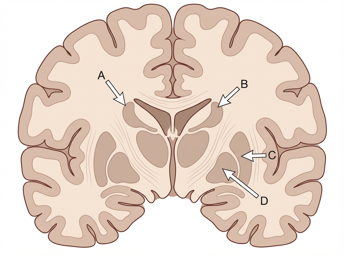
Identify the structure labeled as Caudate nucleus in the given image.

From which structure do the internal arcuate fibres of the medulla originate?
The sternocleidomastoid muscle is supplied by all of the following arteries except?
What is the action of 20mcg/kg/min dopamine?
Which of the following is the largest branch of the vertebral artery?
Which of the following is a DNA repair defect?
What cells form the blood-brain barrier?
What is the recommended maintenance level of mixed venous oxygen saturation in a patient in shock?
From which pharyngeal pouch does the pharyngeal tonsil develop?
Explanation: **Explanation:** **Congo red** is the gold standard histopathological stain for identifying **amyloid**, a pathological proteinaceous substance deposited in tissues. Under a standard light microscope, amyloid fibrils bind to the dye and appear as a **brilliant pink or salmon-pink** color. * **Why Brilliant Pink is correct:** Congo red is a linear molecule that aligns itself parallel to the $\beta$-pleated sheet structure of amyloid fibrils. This specific binding pattern results in the characteristic pinkish-red hue under regular light. * **Why other options are incorrect:** * **Dark brown:** This is characteristic of melanin or hemosiderin (Prussian blue stain is used for the latter). * **Blue:** This is typical of Hematoxylin (staining nuclei) or Alcian blue (staining mucin). * **Khaki:** This is not a standard color description for amyloid staining. **High-Yield Clinical Pearls for NEET-PG:** 1. **Apple-Green Birefringence:** This is the most frequently tested fact. When Congo red-stained amyloid is viewed under **polarized light**, it exhibits a characteristic apple-green birefringence due to the highly organized $\beta$-pleated sheet configuration. 2. **Metachromasia:** Amyloid also shows metachromasia (changing the color of the dye) when stained with **Crystal violet** or **Methyl violet**, appearing rose-pink. 3. **Thioflavin T/S:** These are fluorescent dyes used for amyloid; Thioflavin T produces a bright yellow-green fluorescence. 4. **Precursor Proteins:** Remember that AL (Amyloid Light chain) is associated with Multiple Myeloma, while AA (Amyloid Associated) is seen in chronic inflammatory conditions.
Explanation: ***B*** - The **caudate nucleus** is located in the **lateral ventricle**, specifically forming the lateral wall and floor of the anterior horn of the lateral ventricle. - It has a characteristic **C-shaped** structure with a **head**, **body**, and **tail**, with the head being most prominent in coronal sections at the level of the anterior commissure. *A* - This label likely corresponds to the **putamen**, which is part of the **lentiform nucleus** and lies lateral to the internal capsule. - The putamen is separated from the caudate nucleus by the **anterior limb of the internal capsule** and appears more lateral in brain sections. *C* - This structure is probably the **globus pallidus**, which forms the medial part of the **lentiform nucleus** and lies medial to the putamen. - The globus pallidus appears **paler** on imaging due to its high **myelination** and is involved in **motor control circuits**. *D* - This label most likely indicates the **thalamus**, which is a large **ovoid structure** forming the lateral walls of the third ventricle. - The thalamus lies **medial and posterior** to the caudate nucleus and serves as a major **relay station** for sensory and motor information.
Explanation: The **internal arcuate fibers** are second-order sensory neurons that form a critical part of the **Dorsal Column-Medial Lemniscus (DCML) pathway**, which carries fine touch, vibration, and conscious proprioception. 1. **Why Option D is correct:** First-order neurons from the spinal cord (fasciculus gracilis and cuneatus) synapse in the **nucleus gracilis and nucleus cuneatus** located in the lower medulla. The axons of the second-order neurons emerging from these nuclei sweep anteriorly and medially as the **internal arcuate fibers**. These fibers then **decussate** (cross the midline) in the sensory decussation to form the **medial lemniscus** on the opposite side, which ascends to the thalamus (VPL nucleus). 2. **Why other options are incorrect:** * **Hypoglossal nucleus (A):** This is a motor nucleus (GSE) supplying the muscles of the tongue; its fibers exit the medulla ventrally between the pyramid and olive. * **Dorsal nucleus of vagus (B):** This is a parasympathetic (GVE) nucleus providing autonomic supply to the thorax and abdomen. * **Nucleus tractus solitarius (C):** This is a sensory nucleus for taste (SVA) and visceral sensations (GVA), receiving inputs from cranial nerves VII, IX, and X. **High-Yield Clinical Pearls for NEET-PG:** * **Sensory Decussation:** Occurs at a higher level in the medulla than the motor (pyramidal) decussation. * **Medial Lemniscus Somatotopy:** In the medulla, the medial lemniscus is oriented vertically ("standing man" position), with fibers for the feet (gracilis) located most anteriorly. * **Lesion Localization:** A lesion of the internal arcuate fibers or medial lemniscus results in **contralateral** loss of vibration and position sense.
Explanation: The **sternocleidomastoid (SCM)** is a major landmark muscle of the neck with a segmental and profuse blood supply. Understanding its arterial supply is high-yield for NEET-PG, as it involves branches from both the external carotid and subclavian systems. ### **Why Thyrocervical Trunk is the Correct Answer** The **Thyrocervical trunk** itself does not directly supply the SCM. While its branch, the *suprascapular artery*, may occasionally provide minor twigs to the lower portion of the muscle, the trunk as a whole is not considered a primary supplier. In the context of standard anatomical teaching and competitive exams, the SCM is supplied by direct branches of named arteries rather than the trunk itself. ### **Analysis of Incorrect Options (Suppliers of SCM)** * **Occipital Artery (Option A):** Provides two main branches to the SCM (upper and middle parts). This is a major source of blood supply. * **Posterior Auricular Artery (Option B):** Supplies the uppermost part of the muscle near its insertion on the mastoid process. * **Superior Thyroid Artery (Option D):** Provides a specific "sternocleidomastoid branch" that supplies the middle portion of the muscle. ### **High-Yield Clinical Pearls for NEET-PG** 1. **Nerve Supply:** The SCM has a dual nerve supply: **Spinal Accessory Nerve (CN XI)** for motor function and **C2, C3 ventral rami** for proprioception. 2. **Surgical Landmark:** The SCM divides the neck into anterior and posterior triangles. The **Erb’s point** (nerve point of the neck) is located at the posterior border of the SCM. 3. **Torticollis (Wry neck):** Often caused by fibrosis or hematoma of the SCM, leading to the head tilting toward the affected side and the chin rotating to the opposite side. 4. **Blood Supply Summary:** Remember the mnemonic **"S-O-P-A"** for SCM supply: **S**uperior thyroid, **O**ccipital, **P**osterior auricular, and **A**scending cervical arteries.
Explanation: **Explanation:** The physiological effects of Dopamine are strictly **dose-dependent**, acting on different receptors as the infusion rate increases. This is a high-yield concept for NEET-PG, often referred to as the "Dopamine Dose Ladder." 1. **Why Vasoconstriction is correct:** At high doses (**>10–15 mcg/kg/min**, and specifically at 20 mcg/kg/min), dopamine loses its selectivity for dopaminergic and beta-receptors. It predominantly stimulates **Alpha-1 adrenergic receptors**, leading to potent peripheral vasoconstriction [1]. This increases systemic vascular resistance (SVR). 2. **Analysis of Incorrect Options:** * **Option A (Renal vasodilation):** This occurs at **low doses (0.5–2 mcg/kg/min)** via D1 receptors in the renal vasculature. While it increases renal blood flow, clinical studies have shown it does not prevent acute kidney injury. * **Option D (Increased myocardial contractility):** This occurs at **medium doses (2–10 mcg/kg/min)** via **Beta-1 receptors**. This increases heart rate and stroke volume (inotropic effect). * **Option C (Increased blood pressure):** While high-dose dopamine *does* increase blood pressure, "Vasoconstriction" is the more precise physiological **action** requested by the question. Blood pressure elevation is the *result* of the vasoconstriction and increased cardiac output. **High-Yield Clinical Pearls for NEET-PG:** * **Low Dose (D1):** "Renal dose" (Vasodilation). * **Medium Dose (B1):** "Cardiac dose" (Inotropy). * **High Dose (A1):** "Pressor dose" (Vasoconstriction). * **Antidote:** If extravasation occurs during high-dose infusion, **Phentolamine** (alpha-blocker) is used to prevent tissue necrosis.
Explanation: **Explanation:** The **Posterior Inferior Cerebellar Artery (PICA)** is the largest and most significant branch of the fourth (V4) segment of the vertebral artery. It typically arises near the lower end of the medulla oblongata and follows a tortuous course around the medulla to supply the postero-inferior surface of the cerebellum and the choroid plexus of the fourth ventricle. **Analysis of Options:** * **Option A (Correct):** PICA is the largest branch. It is clinically vital as it supplies the lateral part of the medulla; its occlusion leads to Lateral Medullary Syndrome (Wallenberg Syndrome). * **Option B (Incorrect):** The **Anterior Spinal Artery** is formed by the union of two small branches from the vertebral arteries. While it is long and supplies the anterior two-thirds of the spinal cord, it is smaller in caliber than the PICA. * **Option C (Incorrect):** **Meningeal branches** are small vessels that supply the bone and dura mater of the posterior cranial fossa. They are significantly smaller than the major cerebellar branches. **High-Yield Facts for NEET-PG:** 1. **Origin:** The vertebral artery is the first branch of the first part of the subclavian artery. 2. **Course:** It enters the transverse foramen of the **C6 vertebra** (not C7). 3. **Basilar Artery:** The two vertebral arteries join at the lower border of the pons to form the basilar artery. 4. **Clinical Correlation:** **Wallenberg Syndrome** (PICA occlusion) presents with "crossed anesthesia" (ipsilateral loss of pain/temp on the face and contralateral loss on the body), dysphagia, and ataxia.
Explanation: ### Explanation **Correct Answer: C. Xeroderma pigmentosum** **Mechanism:** Xeroderma pigmentosum (XP) is an autosomal recessive disorder characterized by a defect in **Nucleotide Excision Repair (NER)**. In healthy individuals, the NER pathway identifies and removes pyrimidine dimers (usually thymine dimers) caused by Ultraviolet (UV) radiation. In XP patients, these DNA lesions remain unrepaired, leading to rapid accumulation of mutations, severe photosensitivity, and a 2000-fold increased risk of skin cancers (Basal Cell Carcinoma, Squamous Cell Carcinoma, and Melanoma) at a young age. **Analysis of Incorrect Options:** * **A. Retinoblastoma:** This is caused by a mutation in the **RB1 gene** (a tumor suppressor gene) on chromosome 13q14. It regulates the G1/S checkpoint of the cell cycle, not DNA repair. * **B. Neurofibromatosis:** NF Type 1 is caused by a mutation in the **NF1 gene** (neurofibromin), which acts as a GTPase-activating protein that downregulates the RAS signaling pathway [1]. It is a disorder of cell signaling/growth control. * **D. MEN1 (Multiple Endocrine Neoplasia Type 1):** This is caused by a mutation in the **MEN1 gene** (encoding the protein Menin), which is involved in transcriptional regulation and genome stability, but it is primarily classified as a tumor suppressor gene defect rather than a classic DNA repair pathway defect like XP. **High-Yield NEET-PG Pearls:** * **Other DNA Repair Defects:** * **Lynch Syndrome (HNPCC):** Mismatch Repair (MMR) defect. * **Ataxia-Telangiectasia:** Defect in ATM gene (repair of double-strand DNA breaks). * **Fanconi Anemia:** Defect in homologous recombination/DNA cross-link repair. * **BRCA 1/2:** Defect in homologous recombination repair. * **XP Clinical Triad:** Sun sensitivity, pigmentary changes (freckling), and early-onset cutaneous malignancies.
Explanation: **Explanation:** The **Blood-Brain Barrier (BBB)** is a highly selective semipermeable border that prevents solutes in the circulating blood from non-selectively crossing into the extracellular fluid of the central nervous system (CNS). **Why Astrocytes are correct:** The BBB is structurally composed of three layers: capillary endothelial cells (connected by tight junctions), a thick basement membrane, and the **foot processes of Astrocytes** (perivascular end-feet). Astrocytes play a critical role by inducing and maintaining the tight junctions between endothelial cells, thereby regulating the transport of nutrients and waste [1]. **Analysis of Incorrect Options:** * **A. Microglia:** These are the resident macrophages of the CNS [1]. They act as the primary immune defense and are derived from the mesoderm (monocyte-macrophage lineage), not the neuroectoderm [1]. * **B. Oligodendrocytes:** These cells are responsible for the myelination of axons within the CNS (analogous to Schwann cells in the PNS) [3]. Each oligodendrocyte myelinates numerous internodes on multiple axons [3]. * **D. Type II Pneumocytes:** These are cells found in the alveoli of the lungs; they secrete pulmonary surfactant to reduce surface tension. **High-Yield Clinical Pearls for NEET-PG:** * **Tight Junctions (Zonula occludens):** These are the most important physiological component of the BBB. * **Circumventricular Organs (CVOs):** These are specific areas where the BBB is absent (e.g., Area Postrema, Neurohypophysis, Pineal gland), allowing the brain to monitor systemic chemical changes [2]. * **Clinical Relevance:** Non-polar/lipid-soluble substances (e.g., CO2, O2, alcohol) cross the BBB easily, while large polar molecules (e.g., most antibiotics) do not [4]. This is why drugs like **L-Dopa** are used for Parkinson’s instead of Dopamine, as Dopamine cannot cross the BBB.
Explanation: The management of shock revolves around ensuring adequate oxygen delivery ($DO_2$) to meet cellular oxygen demand ($VO_2$). **Mixed Venous Oxygen Saturation ($SvO_2$)** is a crucial hemodynamic parameter measured from the pulmonary artery that reflects the balance between systemic oxygen delivery and consumption [1]. 1. **Why 50-70% is Correct:** In a healthy resting individual, $SvO_2$ is typically **65-75%**. In states of shock, tissues compensate for decreased delivery by increasing oxygen extraction. A maintenance level of **50-70%** is considered the "therapeutic window" in clinical practice. Maintaining $SvO_2$ above 50% (specifically aiming for >65-70% in early goal-directed therapy) ensures that the body is not excessively exhausting its venous oxygen reserve, thereby preventing anaerobic metabolism and lactic acidosis [2]. 2. **Analysis of Incorrect Options:** * **>70% (Option A):** While ideal in healthy individuals, maintaining >70% in a shock patient can sometimes indicate "shunting" or a failure of tissues to extract oxygen (e.g., in late-stage septic shock), or it may represent an unnecessarily high-pressure resuscitation [2]. * **40-50% (Option C):** This range indicates significant physiological stress and inadequate oxygen delivery. It suggests that the compensatory extraction is reaching its limit. * **<40% (Option D):** This is a critical level. When $SvO_2$ falls below 40%, the compensatory mechanisms fail, leading to profound tissue hypoxia, a shift to anaerobic metabolism, and rising serum lactate. **High-Yield Clinical Pearls for NEET-PG:** * **Gold Standard Measurement:** $SvO_2$ is measured via a **Swan-Ganz (Pulmonary Artery) catheter**. * **$ScvO_2$ vs. $SvO_2$:** Central Venous Oxygen Saturation ($ScvO_2$), measured from the SVC, is usually **5-10% higher** than $SvO_2$ because it does not include the highly deoxygenated blood from the coronary sinus [2]. * **Low $SvO_2$ causes:** Decreased Cardiac Output (CO), decreased $Hb$, decreased $SaO_2$, or increased $VO_2$ (shivering/fever). * **High $SvO_2$ causes:** Sepsis (maldistribution of flow), cyanide poisoning (histotoxic hypoxia), or left-to-right shunts.
Explanation: The development of the pharyngeal apparatus is a high-yield topic for NEET-PG. Here is the breakdown of the pharyngeal pouch derivatives: **Why the Correct Answer (B) is Right:** The **Second Pharyngeal Pouch** is responsible for the development of the **palatine tonsil**. The endodermal lining of this pouch proliferates to form buds that penetrate the surrounding mesoderm. These buds are later infiltrated by lymphatic tissue to form the definitive tonsil. While the question uses the term "pharyngeal tonsil" (which is technically the adenoid located in the nasopharynx), in the context of standard medical examinations and embryological pouch derivatives, the **palatine tonsil** is the primary derivative of the second pouch. **Why the Other Options are Wrong:** * **Option A (First Pouch):** This pouch forms the **tubotympanic recess**, which gives rise to the epithelial lining of the auditory (Eustachian) tube and the middle ear cavity (tympanic cavity). * **Option C (Third Pouch):** This pouch has dorsal and ventral wings. The dorsal part forms the **inferior parathyroid glands**, and the ventral part forms the **thymus**. * **Option D (Fourth Pouch):** The dorsal part forms the **superior parathyroid glands**, while the ventral part (often associated with the 5th/6th pouch) forms the **ultimobranchial body**, which gives rise to the parafollicular (C) cells of the thyroid. **High-Yield Clinical Pearls for NEET-PG:** * **Rule of "3-4":** The 3rd pouch forms the *inferior* parathyroid, and the 4th pouch forms the *superior* parathyroid. The 3rd pouch derivatives migrate further caudally. * **DiGeorge Syndrome:** Results from the failure of the 3rd and 4th pouches to develop, leading to thymic hypoplasia (immunodeficiency) and hypocalcemia (absent parathyroids). * **Tonsillar Fossa:** The remains of the second pharyngeal pouch persist in the adult as the supratonsillar fossa.
Organization of the Nervous System
Practice Questions
Spinal Cord Anatomy
Practice Questions
Brainstem Anatomy
Practice Questions
Cerebellum
Practice Questions
Diencephalon
Practice Questions
Cerebral Cortex
Practice Questions
Basal Ganglia
Practice Questions
Limbic System
Practice Questions
Cranial Nerves
Practice Questions
Autonomic Nervous System
Practice Questions
Neural Pathways and Tracts
Practice Questions
Neurovascular Anatomy
Practice Questions
Get full access to all questions, explanations, and performance tracking.
Start For Free