
Neuroanatomy — MCQs

On this page
Which structure is located at the floor of the 4th ventricle?
In trochlear palsy, which specific eye movement is lost?

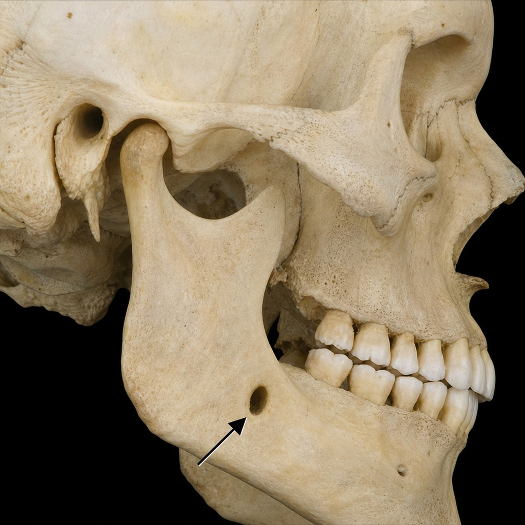
Which nerve passes through the marked foramen in the image?
Contralateral loss of pain and temperature is due to injury to:
Which of the following is NOT part of the special visceral efferent column?
Traumatic optic neuropathy due to closed head trauma commonly affects which part of the optic nerve?
Pupillary reflex pathway - All of the following are a part except?
Myelination in peripheral nervous system is done by
Which of the following is pure sensory nerve?
Largest cranial nerve is:
Practice by Chapter
Organization of the Nervous System
Practice Questions
Spinal Cord Anatomy
Practice Questions
Brainstem Anatomy
Practice Questions
Cerebellum
Practice Questions
Diencephalon
Practice Questions
Cerebral Cortex
Practice Questions
Basal Ganglia
Practice Questions
Limbic System
Practice Questions
Cranial Nerves
Practice Questions
Autonomic Nervous System
Practice Questions
Neural Pathways and Tracts
Practice Questions
Neurovascular Anatomy
Practice Questions
Want unlimited practice?
Get full access to all questions, explanations, and performance tracking.
Scan to download app