
Neuroanatomy — MCQs

On this page
A 34-year-old male complains of hyperacusis (sensitivity to loud sounds). Injury to which of the following cranial nerves is responsible?
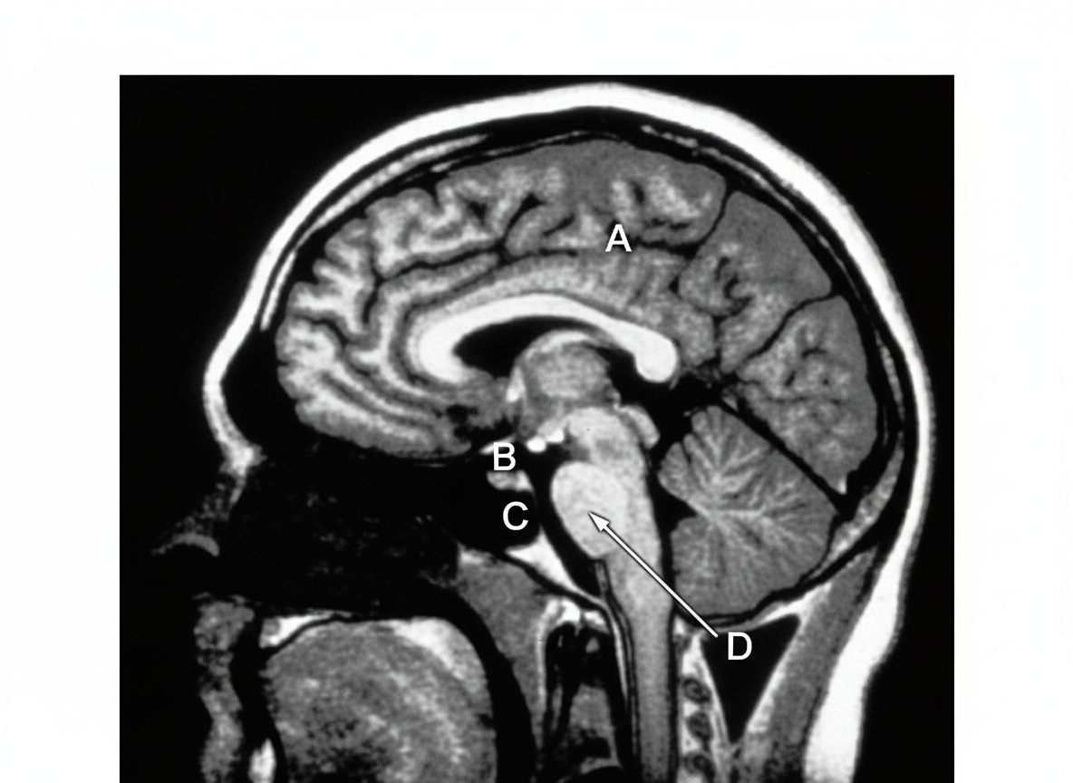
T1 weighted MRI of midsagittal section of the brain is shown. Which among the marked structures contains the two lateral foramina of Luschka?
All the following structures have a Blood-Brain Barrier except?
A 'blow-out fracture' of the orbit most commonly involves which part?
Which of the following is a WRONG pair regarding features of cranial nerves?
Which cranial nerve is the only one that innervates a muscle contralaterally?
Which cranial nerve's fibers are myelinated by oligodendrocytes?
All of the following are true about the facial colliculus EXCEPT:
Hemiplegia is commonly associated with infarction of the area of distribution of which artery?
All of the following are features of ischemia in the anterior choroidal artery territory except?
Practice by Chapter
Organization of the Nervous System
Practice Questions
Spinal Cord Anatomy
Practice Questions
Brainstem Anatomy
Practice Questions
Cerebellum
Practice Questions
Diencephalon
Practice Questions
Cerebral Cortex
Practice Questions
Basal Ganglia
Practice Questions
Limbic System
Practice Questions
Cranial Nerves
Practice Questions
Autonomic Nervous System
Practice Questions
Neural Pathways and Tracts
Practice Questions
Neurovascular Anatomy
Practice Questions
Want unlimited practice?
Get full access to all questions, explanations, and performance tracking.
Scan to download app