
Neuroanatomy — MCQs

On this page
The medulla oblongata receives its blood supply from all of the following arteries except:
Nucleus tractus solitarius receives fibers from all of the following cranial nerves EXCEPT:
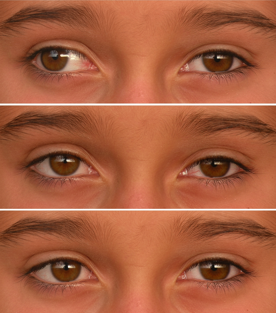
Which cranial nerve is involved in the clinical presentation shown below?
A 5-day-old infant male presents with an abnormally large head. A CT scan reveals enlarged lateral and third ventricles with a normal-sized fourth ventricle, suggesting stenosis of the cerebral aqueduct (of Sylvius). Which of the following conditions is characteristic of these symptoms?
Which cranial nerve is most commonly involved in posterior communicating artery aneurysm?
Which of the following statements is NOT true regarding the 5th cranial nerve?
Which of the following vessels do not take part in the Circle of Willis?
Which of the following is NOT a consequence of III nerve palsy?
The bulbocavernosus reflex is elicited by stimulation of which of the following?
All of the following are found in the floor of the third ventricle, except:
Practice by Chapter
Organization of the Nervous System
Practice Questions
Spinal Cord Anatomy
Practice Questions
Brainstem Anatomy
Practice Questions
Cerebellum
Practice Questions
Diencephalon
Practice Questions
Cerebral Cortex
Practice Questions
Basal Ganglia
Practice Questions
Limbic System
Practice Questions
Cranial Nerves
Practice Questions
Autonomic Nervous System
Practice Questions
Neural Pathways and Tracts
Practice Questions
Neurovascular Anatomy
Practice Questions
Want unlimited practice?
Get full access to all questions, explanations, and performance tracking.
Scan to download app