
Neuroanatomy — MCQs

On this page
The hypoglossal nerve is primarily what type of nerve?
Injury to which of the following structures causes constricted pupils even when room lighting is dim?
The ventricles of the brain are lined by which type of cells?
In a newborn, at which vertebral level does the spinal cord typically end?
Injury to the cervical sympathetic trunk produces Horner's syndrome. Which of the following is NOT a characteristic sign of Horner's syndrome?
All of the following arise from the occulomotor nerve except?
Which nucleus of the cerebellum is primarily responsible for controlling slow pursuit and saccadic eye movements?
The lentiform nucleus of the basal ganglia includes which of the following structures?
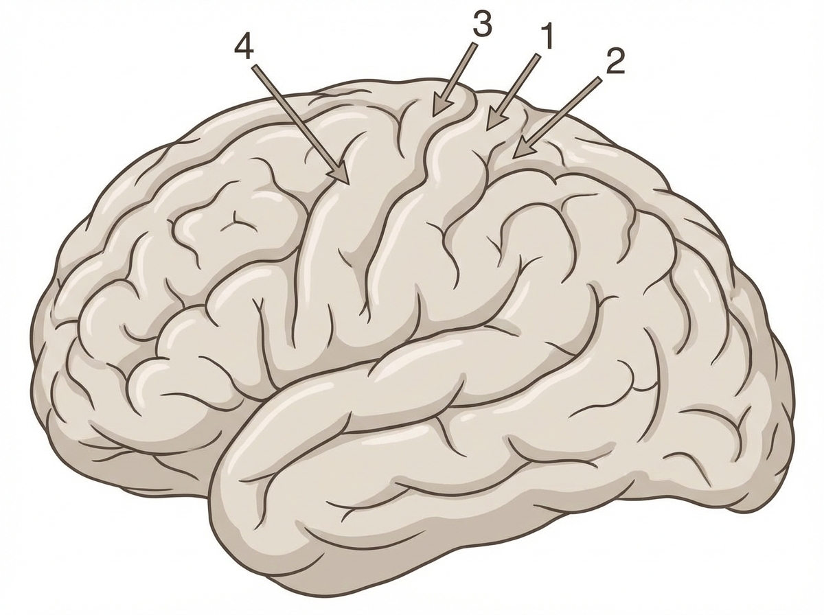
Which of the following areas of the brain is not a primary sensory cortex?
Which of the following statements regarding Arnold-Chiari malformation is INCORRECT?
Practice by Chapter
Organization of the Nervous System
Practice Questions
Spinal Cord Anatomy
Practice Questions
Brainstem Anatomy
Practice Questions
Cerebellum
Practice Questions
Diencephalon
Practice Questions
Cerebral Cortex
Practice Questions
Basal Ganglia
Practice Questions
Limbic System
Practice Questions
Cranial Nerves
Practice Questions
Autonomic Nervous System
Practice Questions
Neural Pathways and Tracts
Practice Questions
Neurovascular Anatomy
Practice Questions
Want unlimited practice?
Get full access to all questions, explanations, and performance tracking.
Scan to download app