
Neuroanatomy — MCQs

On this page
Which of the following is not a component of the medial reticular column?
The vestibulocerebellar tract terminates in which part of the cerebellum?
Cyclosporine is an:
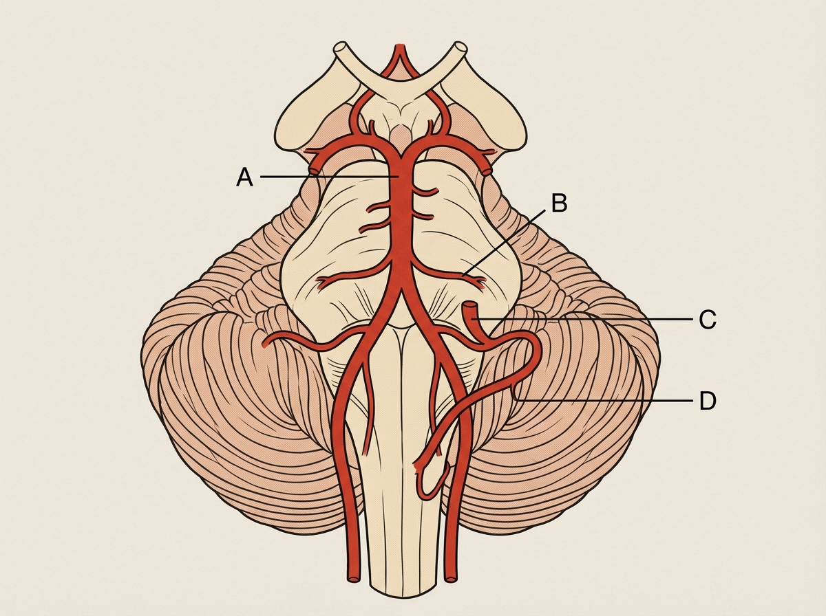
The anterior inferior cerebellar artery is a branch of which artery?
The cranial nerve nucleus supplying the marked muscle lies at the level of:

Transection of the anterolateral spinothalamic tract results in what kind of sensory deficit?
In which nucleus does the dorsal column relay occur?
All of the following cranial nerves contain somatic efferents except?
Which artery is involved in Wallenberg syndrome?
Wallenberg's syndrome does not involve which cranial nerve?
Practice by Chapter
Organization of the Nervous System
Practice Questions
Spinal Cord Anatomy
Practice Questions
Brainstem Anatomy
Practice Questions
Cerebellum
Practice Questions
Diencephalon
Practice Questions
Cerebral Cortex
Practice Questions
Basal Ganglia
Practice Questions
Limbic System
Practice Questions
Cranial Nerves
Practice Questions
Autonomic Nervous System
Practice Questions
Neural Pathways and Tracts
Practice Questions
Neurovascular Anatomy
Practice Questions
Want unlimited practice?
Get full access to all questions, explanations, and performance tracking.
Scan to download app