A patient exhibiting signs of Horner's syndrome likely has a lesion in which part of the sympathetic nervous system?
Which nerve innervates the muscles of mastication?
A patient presents with an inability to abduct the eye. Which cranial nerve is most likely affected?
A 50-year-old male presents with unilateral ptosis, miosis, and anhidrosis. Which structure is most likely involved?
A neurologist is evaluating a patient with an altered sense of taste. Damage to which cranial nerve is most likely responsible for this symptom?
A patient experiencing vertigo and tinnitus may have pathology in which cranial nerve?
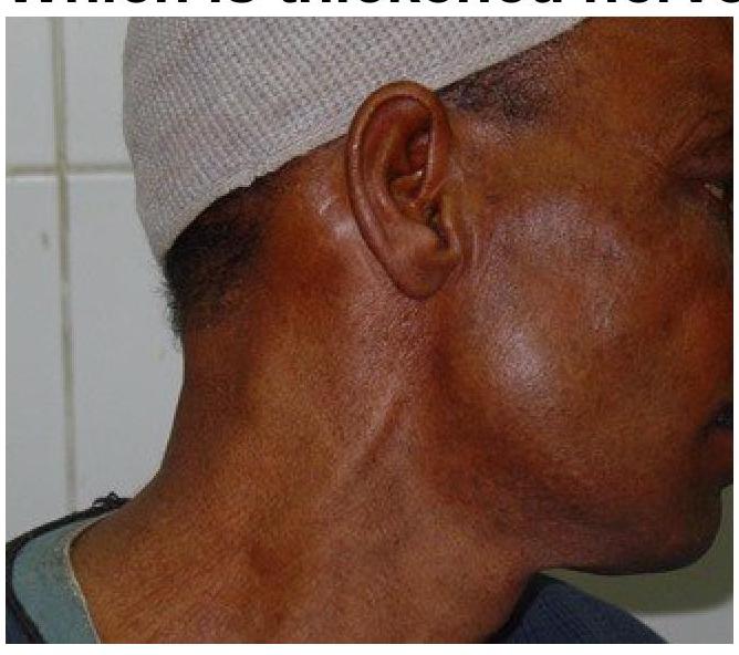
Which thickened nerve is shown in the image?

Which of the following is NOT a special visceral efferent (SVE) nerve?
In which part of the inner ear are cristae located?
HSV-2 causes latent infection in which nerve plexus/ ganglia ?
Explanation: ***Cervical ganglia*** - **Horner's syndrome** results from interruption of the **sympathetic pathway** to the eye, which crucially involves the **superior cervical ganglion** in the neck [1]. - This ganglion is part of the **cervical sympathetic chain** and provides **postganglionic fibers** that innervate the dilator pupillae muscle, the superior tarsal muscle (Müller's muscle), and facial sweat glands. - Lesions affecting the **cervical sympathetic trunk** or the **superior cervical ganglion** itself are the most direct anatomical correlates of Horner's syndrome. *Thoracic ganglia* - While **preganglionic sympathetic fibers** for the oculosympathetic pathway originate from **T1-T3 spinal segments** [1] and ascend through the sympathetic chain, the question asks about the part of the sympathetic nervous system most directly associated with Horner's syndrome. - Lesions at the **thoracic level** (such as Pancoast tumors at the lung apex) can cause Horner's syndrome by affecting preganglionic fibers, but the **cervical sympathetic pathway** remains the most specific anatomical answer. - The **thoracic ganglia** primarily serve the thoracic and abdominal viscera. *Lumbar ganglia* - **Lumbar ganglia** provide sympathetic innervation to the **lower abdomen** and **lower limbs**. - They have no role in the sympathetic pathway to the head and neck or in Horner's syndrome. *Sacral ganglia* - The **sacral ganglia** contribute to sympathetic innervation of **pelvic organs**. - They have no connection to the oculosympathetic pathway or the presentation of Horner's syndrome.
Explanation: ***Trigeminal nerve*** - The **trigeminal nerve (CN V)** is responsible for both sensory innervation of the face and motor innervation of the **muscles of mastication**. - The **mandibular division (V3)** of the trigeminal nerve specifically carries the motor fibers to these muscles. *Facial nerve* - The **facial nerve (CN VII)** primarily controls the **muscles of facial expression**, not mastication. - It also provides taste sensation to the anterior two-thirds of the tongue and innervates various glands. *Glossopharyngeal nerve* - The **glossopharyngeal nerve (CN IX)** is primarily involved in **swallowing**, taste sensation to the posterior tongue, and innervating the parotid gland. - It does not have a direct role in controlling the muscles used for chewing. *Hypoglossal nerve* - The **hypoglossal nerve (CN XII)** provides motor innervation to almost all of the **intrinsic and extrinsic muscles of the tongue**. - Its primary function is in tongue movement for speech and swallowing, not mastication.
Explanation: Correct: Abducens nerve - The abducens nerve (CN VI) specifically innervates the lateral rectus muscle of the eye - The lateral rectus muscle is responsible for abduction (moving the eye laterally/outwards) [1] - Damage to CN VI results in the inability to abduct the affected eye, causing horizontal diplopia (double vision) that worsens when looking toward the affected side Incorrect: Oculomotor nerve - The oculomotor nerve (CN III) innervates most extrinsic eye muscles (medial rectus, superior rectus, inferior rectus, and inferior oblique), but NOT the lateral rectus [1] - Damage to CN III typically causes ptosis, diplopia, and an eye that is positioned down and out at rest - Would cause inability to ADduct, elevate, or depress the eye, not ABduct [1] Incorrect: Trochlear nerve - The trochlear nerve (CN IV) innervates only the superior oblique muscle - Superior oblique is responsible for intorsion, depression (especially when the eye is adducted), and slight abduction [1] - Injury causes vertical diplopia that worsens when looking down and inward (e.g., reading or descending stairs) Incorrect: Optic nerve - The optic nerve (CN II) is a sensory nerve responsible for vision and transmitting visual information to the brain - It has NO role in eye movement - Damage causes visual loss, field defects, or blindness, not motor deficits
Explanation: ***Superior cervical ganglion*** - This symptom complex of **ptosis**, **miosis**, and **anhidrosis** on one side of the face is known as **Horner's syndrome** [1]. - **Horner's syndrome** results from a lesion affecting the **sympathetic pathway** to the eye and face, which involves synapses in the superior cervical ganglion [1]. - The postganglionic sympathetic fibers from this ganglion innervate the **dilator pupillae muscle**, **Müller's muscle** (upper eyelid), and **facial sweat glands** [1]. *Ciliary ganglion* - The ciliary ganglion is a **parasympathetic ganglion** involved in innervation to the eye, providing fibers that control **pupil constriction** (via sphincter pupillae) and **accommodation** [2]. - A lesion here would cause **loss of parasympathetic function**, resulting in a **dilated pupil** (mydriasis) due to unopposed sympathetic activity and loss of accommodation, but not ptosis or anhidrosis [2]. *Trigeminal ganglion* - The trigeminal ganglion contains cell bodies for **sensory innervation** to the face. - Lesions of the trigeminal ganglion would present with **facial numbness** or **pain**, not the classic triad of Horner's syndrome. *Otic ganglion* - The otic ganglion is a **parasympathetic ganglion** primarily involved in the innervation of the **parotid gland**, controlling **salivation**. - Dysfunction of the otic ganglion would affect salivary production, but not ocular symptoms or facial sweating.
Explanation: ***Facial nerve*** - The **facial nerve (CN VII)** carries taste sensation from the **anterior two-thirds of the tongue** via the **chorda tympani branch** [1]. - Damage to this nerve, for example, in conditions like **Bell's palsy**, can lead to an altered sense of taste (ageusia or dysgeusia) [1]. - CN VII is the **most commonly affected** cranial nerve causing taste disturbances in clinical practice and is the **primary nerve tested** for taste function [2]. - Note: The **glossopharyngeal nerve (CN IX)** also carries taste from the posterior one-third of the tongue, but CN VII covers a larger area and is more frequently affected in clinical conditions. *Hypoglossal nerve* - The **hypoglossal nerve (CN XII)** is primarily responsible for the **motor control of the tongue muscles**, enabling speech and swallowing. - It does **not carry sensory information** related to taste, only motor function. *Vagus nerve* - The **vagus nerve (CN X)** has a **minor role** in taste sensation from the **epiglottis and posterior pharynx**, which represents a very small area [1]. - This minimal contribution is **not typically described** as causing a general altered sense of taste. - Its main functions include parasympathetic innervation to the thoracic and abdominal viscera. *Trigeminal nerve* - The **trigeminal nerve (CN V)** is responsible for **general sensation to the face** and head, and motor control of the muscles of mastication. - It does **not carry taste sensation** (special sensory), although it does convey **general sensation** (touch, temperature, pain) from the tongue.
Explanation: ***Vestibulocochlear nerve*** - The **vestibular portion** of this nerve is responsible for balance, and its dysfunction can lead to **vertigo** [1]. - The **cochlear portion** transmits auditory information, and its pathology causes **tinnitus** (ringing in the ears) [2]. *Hypoglossal nerve* - This nerve is primarily involved in controlling the muscles of the **tongue** for movement and speech. - Dysfunction typically results in **dysarthria** and **deviation of the tongue**, not vertigo or tinnitus. *Facial nerve* - The facial nerve controls muscles of **facial expression**, taste perception in the anterior two-thirds of the tongue, and tear/salivary gland function. - Pathology often presents as **facial paralysis** (e.g., Bell's palsy) or altered taste, not issues related to balance or hearing. *Glossopharyngeal nerve* - This nerve is responsible for taste in the posterior third of the tongue, sensation from the pharynx, and control of the **stylopharyngeus muscle** for swallowing. - Damage can lead to **dysphagia**, loss of gag reflex, or altered taste, none of which include vertigo or tinnitus.
Explanation: ***Greater auricular nerve*** - The image clearly shows a **thickened, rope-like structure** running superficially on the side of the neck, ascending towards the earlobe. - This anatomical location and appearance are highly characteristic of an enlarged **greater auricular nerve**, often seen in conditions like **leprosy**. *Facial Nerve* - The **facial nerve** is primarily a motor nerve that innervates the muscles of facial expression and is located deeper within the parotid gland. - It would not typically present as a thickened, superficial structure visible on the surface of the neck or behind the earlobe. *Vagus Nerve* - The **vagus nerve** is a cranial nerve with extensive autonomic functions, running through the neck within the carotid sheath, much deeper than the structure shown. - It is not superficially visible or palpable in this manner under normal or pathological conditions that cause thickening. *Glossopharyngeal Nerve* - The **glossopharyngeal nerve** is another cranial nerve that exits the skull and descends in the neck, primarily involved in swallowing and taste. - Like the vagus nerve, it is located deep within the neck and would not be visible or thickened superficially as depicted.
Explanation: ***Nucleus ambiguus*** - The **nucleus ambiguus** is a **brainstem nucleus** that contains the cell bodies of motor neurons, making it a source of SVE fibers, but it is not a nerve itself. - It provides motor innervation to muscles of the **pharynx, larynx, and soft palate** via cranial nerves IX, X, and XI. *Glossopharyngeal n* - The **glossopharyngeal nerve (CN IX)** is a **mixed nerve** that contains efferent (motor) fibers, including special visceral efferent (SVE) components. - Its SVE fibers originate from the **nucleus ambiguus** and innervate the **stylopharyngeus muscle**, involved in swallowing. *vagus nerve* - The **vagus nerve (CN X)** is a **mixed cranial nerve** with significant special visceral efferent (SVE) functions. - Its SVE fibers originate from the **nucleus ambiguus** and innervate the muscles of the **pharynx, larynx, and soft palate**, crucial for speech and swallowing. *trigeminal nerve* - The **trigeminal nerve (CN V)** contains a **mandibular division (V3)** that includes special visceral efferent (SVE) fibers. - These SVE fibers originate from the **motor nucleus of the trigeminal nerve** and innervate the **muscles of mastication**.
Explanation: - **Cristae ampullares** are sensory organs found within the **ampullae** of the semicircular canals [1]. - They are responsible for detecting **angular acceleration** (rotational movements) of the head [1]. *Utricle* - The utricle contains **maculae**, not cristae, which detect **linear acceleration** and the position of the head relative to gravity [1]. - Its sensory hair cells are covered by an **otolithic membrane** [1]. *Saccule* - Similar to the utricle, the saccule also contains **maculae** for detecting **linear acceleration** and vertical head movements [1]. - It plays a role in sensing gravity and vertical motion [1]. *Otolith membrane* - The **otolith membrane** is a gel-like structure embedded with **otoliths (calcium carbonate crystals)** that overlie the hair cells in the maculae of the utricle and saccule [1]. - This membrane is not a location for cristae, but rather a component of the sensory mechanism for **linear acceleration** and gravity.
Explanation: ***Sacral ganglia*** * **HSV-2** primarily causes **genital herpes**, and the virus establishes **latency** in the sensory neurons of the sacral ganglia, which innervate the genital area [1]. * During reactivation, the virus travels down these nerves to cause recurrent lesions in the anogenital region. *Trigeminal ganglia* * The **trigeminal ganglia** are the primary site of latency for **HSV-1**, which is associated with **oral herpes** (cold sores) [1]. * Reactivation from the trigeminal ganglia leads to lesions typically around the mouth. *Otic ganglia* * The **otic ganglia** are **parasympathetic ganglia** associated with the glossopharyngeal nerve and are involved in salivary gland function, not HSV latency. * They are not known to be a site of latency for herpes simplex viruses. *Ciliary ganglia* * The **ciliary ganglia** are also **parasympathetic ganglia** associated with the oculomotor nerve, involved in controlling intrinsic eye muscles (e.g., pupillary constriction). * They do not serve as a site for HSV latency.
Organization of the Nervous System
Practice Questions
Spinal Cord Anatomy
Practice Questions
Brainstem Anatomy
Practice Questions
Cerebellum
Practice Questions
Diencephalon
Practice Questions
Cerebral Cortex
Practice Questions
Basal Ganglia
Practice Questions
Limbic System
Practice Questions
Cranial Nerves
Practice Questions
Autonomic Nervous System
Practice Questions
Neural Pathways and Tracts
Practice Questions
Neurovascular Anatomy
Practice Questions
Get full access to all questions, explanations, and performance tracking.
Start For Free