Which nucleus in the brain is common to the IX, X, and XI cranial nerves?
General visceral fibres do not supply which of the following structures?
Which of the following is NOT a branch of the intracranial part of the Internal carotid artery?
Paradoxical splitting of the second heart sound is heard in which of the following conditions?
Cell matrix adhesion are mediated by?
How many nuclei does the trigeminal nerve have?
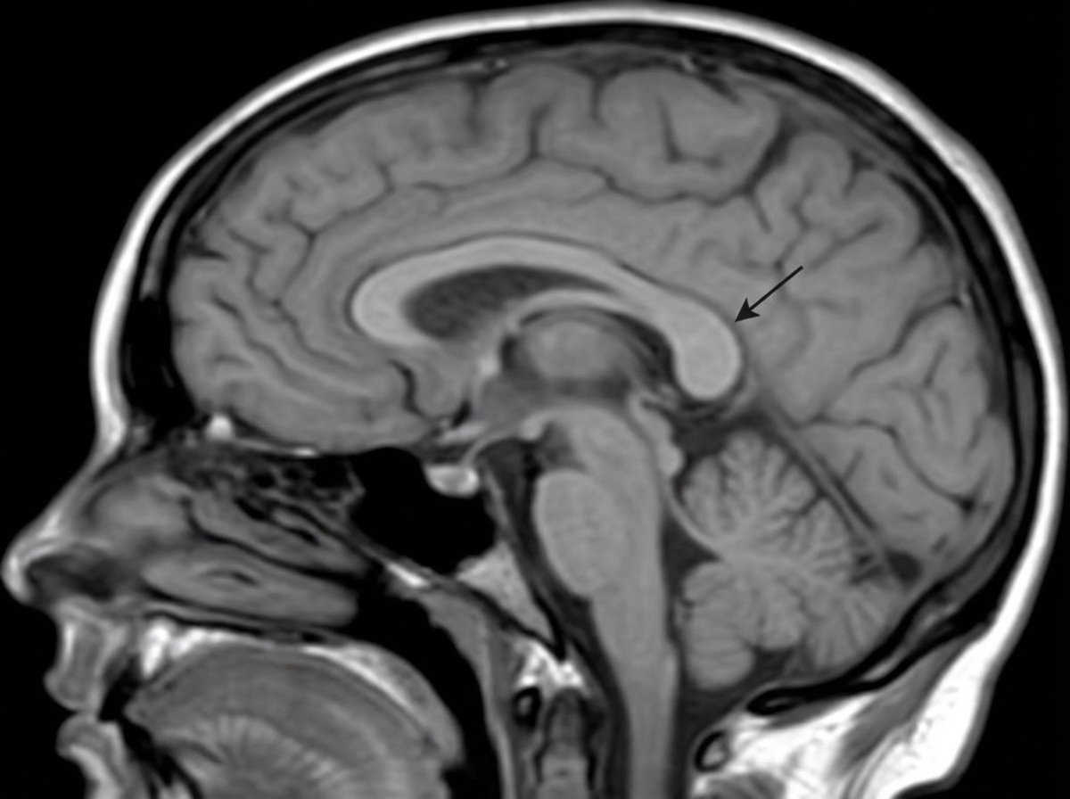
Identify the structure indicated by the arrow in the corpus callosum.

Lateral Medullary Syndrome involves all of the following cranial nerves, EXCEPT:
Which is the most prominent spinous process?
An apical lung tumor can cause which of the following signs?
Explanation: The **Nucleus Ambiguus** is a motor nucleus located in the medulla oblongata. It contains the cell bodies of lower motor neurons that provide **Special Visceral Efferent (SVE)** fibers to the muscles of the pharynx, larynx, and soft palate [1]. ### Why Nucleus Ambiguus is Correct: It serves as the common motor nucleus for three cranial nerves: * **Glossopharyngeal (IX):** Supplies the stylopharyngeus muscle. * **Vagus (X):** Supplies the muscles of the pharynx, soft palate, and larynx. * **Cranial accessory (XI):** Its fibers join the vagus nerve to supply the laryngeal muscles. ### Why Other Options are Incorrect: * **Nucleus Solitarius:** This is a **sensory** nucleus. It receives taste (SVA) from VII, IX, and X, and general visceral sensations (GVA) from IX and X. It is not a motor nucleus. * **Dentate Nucleus:** This is the largest of the deep cerebellar nuclei involved in the planning and initiation of voluntary movements. It has no direct connection to the IX, X, or XI cranial nerves. * **Red Nucleus:** Located in the midbrain, it is part of the extrapyramidal system (rubrospinal tract) involved in motor coordination, primarily of the upper limbs. ### High-Yield Clinical Pearls for NEET-PG: * **Lesion of Nucleus Ambiguus:** Results in nasal regurgitation of fluids, dysphagia (difficulty swallowing), and dysphonia (hoarseness of voice) due to paralysis of the laryngeal and pharyngeal muscles. * **Wallenberg Syndrome (PICA Syndrome):** The nucleus ambiguus is characteristically involved in Lateral Medullary Syndrome, leading to ipsilateral vocal cord paralysis and loss of gag reflex. * **Mnemonic:** Remember **"Ambiguus = Motor"** (Muscles) and **"Solitarius = Sensory"** (Sensation/Taste).
Explanation: **Explanation:** The classification of nerve fibers in neuroanatomy is based on the structures they innervate. The term **General Visceral Efferent (GVE)** refers to the autonomic nervous system fibers that provide motor supply to involuntary structures [2]. 1. **Why Skeletal Muscle is the correct answer:** Skeletal muscles are derived from somites (myotomes) and are under voluntary control. They are supplied by **General Somatic Efferent (GSE)** fibers (e.g., alpha motor neurons in the ventral horn of the spinal cord) [2]. Since GVE fibers only target involuntary "visceral" structures, they do not supply skeletal muscles. 2. **Analysis of Incorrect Options:** * **Smooth Muscles (A):** These are involuntary muscles found in the walls of blood vessels and hollow organs. They are a primary target of GVE (autonomic) fibers [1], [3]. * **Cardiac Muscles (C):** The specialized muscle of the heart is involuntary and regulated by the autonomic nervous system via GVE fibers. * **Glands (D):** Secretomotor supply to sweat glands, salivary glands, and lacrimal glands is provided by GVE fibers (Sympathetic and Parasympathetic). **High-Yield Clinical Pearls for NEET-PG:** * **GVE (General Visceral Efferent):** Think "Autonomic." These are the pre- and post-ganglionic fibers of the sympathetic and parasympathetic systems [4]. * **SVE (Special Visceral Efferent):** These fibers supply muscles derived from the **pharyngeal (branchial) arches**, such as the muscles of mastication and facial expression. Despite being skeletal muscles, they are classified as "visceral" due to their embryological origin. * **GSE (General Somatic Efferent):** Supplies all skeletal muscles *except* those derived from branchial arches (e.g., limb muscles, extraocular muscles, and tongue muscles).
Explanation: ### Explanation The **Internal Carotid Artery (ICA)** enters the cranial cavity and terminates by dividing into its major branches. To answer this question, one must distinguish between the direct branches of the ICA and the components of the **Circle of Willis**. **1. Why "Anterior communicating artery" is the correct answer:** The **Anterior communicating artery** is a short vessel that connects the two Anterior Cerebral Arteries (ACA). While it is a vital part of the Circle of Willis, it is **not** a direct branch of the ICA itself [1]. It arises from the ACAs to complete the anterior portion of the arterial circle. **2. Analysis of Incorrect Options:** * **Anterior cerebral artery (ACA):** This is one of the two terminal branches of the ICA. It supplies the medial surface of the cerebral hemispheres. * **Middle cerebral artery (MCA):** This is the larger terminal branch of the ICA and is often considered its direct continuation. It supplies the majority of the lateral convexity of the brain. * **Posterior communicating artery (PCoA):** This is a pre-terminal branch of the intracranial (cerebral) part of the ICA. It connects the ICA system to the Vertebro-basilar system (specifically the Posterior Cerebral Artery) [1]. **3. Clinical Pearls & High-Yield Facts for NEET-PG:** * **Mnemonic for ICA branches (Cerebral part):** **"O P A M"** — **O**phthalmic, **P**osterior communicating, **A**nterior choroidal, and the terminal branches: **A**nterior cerebral and **M**iddle cerebral arteries. * **Berry Aneurysms:** The most common site for a Berry aneurysm is the junction of the **Anterior communicating artery** and the ACA. * **Stroke Correlation:** The MCA is the most common artery involved in ischemic strokes; the ICA provides roughly 80% of the brain's blood supply [1].
Explanation: The second heart sound (S2) consists of two components: **A2 (Aortic)** and **P2 (Pulmonary)**. Normally, A2 precedes P2, and the gap increases during inspiration (Physiological Splitting) [1]. **Paradoxical (Reversed) Splitting** occurs when A2 is significantly delayed, causing it to occur *after* P2. **Why LBBB is the Correct Answer:** In **Left Bundle Branch Block (LBBB)**, there is delayed depolarization of the left ventricle. This results in delayed closure of the aortic valve (A2). Consequently, P2 occurs before A2. During inspiration, the normal delay of P2 moves it closer to the delayed A2, narrowing the split. During expiration, P2 occurs earlier, widening the gap between P2 and A2. This "reverse" behavior compared to normal physiology defines paradoxical splitting. **Analysis of Incorrect Options:** * **RBBB (Option A):** Causes delayed right ventricular contraction, further delaying P2. This leads to **Wide Physiological Splitting** (A2 and P2 are far apart, widening further on inspiration). * **ASD (Option B):** Characterized by **Fixed Splitting**. The constant volume overload of the right heart keeps the P2 delay constant, regardless of the respiratory cycle. * **VSD (Option C):** Typically causes **Wide Physiological Splitting** due to increased right-sided volume and delayed P2, or it may be associated with a loud single S2 if pulmonary hypertension develops (Eisenmenger syndrome). **NEET-PG High-Yield Pearls:** * **Paradoxical Splitting Causes:** LBBB, Aortic Stenosis (severe), HOCM, and Patent Ductus Aueriosus (PDA). * **Fixed Splitting:** Pathognomonic for Atrial Septal Defect (ASD). * **Wide Splitting:** RBBB, Pulmonary Stenosis, and Mitral Regurgitation (due to early A2). * **Mnemonic for Paradoxical Split:** "The **L**eft side is **L**ate" (**L**BBB, **A**ortic **S**tenosis).
Explanation: **Explanation:** Cell adhesion molecules (CAMs) are essential proteins that facilitate interactions between cells and their environment. They are broadly categorized into two types: those mediating **cell-cell** adhesion and those mediating **cell-matrix** adhesion. **Why Integrins are correct:** Integrins are transmembrane heterodimers (composed of $\alpha$ and $\beta$ subunits) that primarily function as receptors for the **extracellular matrix (ECM)**. They bind to components like fibronectin, laminin, and collagen. By linking the ECM to the intracellular actin cytoskeleton, integrins play a crucial role in signal transduction and structural stability (e.g., in hemidesmosomes) [1]. **Analysis of Incorrect Options:** * **Cadherins:** These are calcium-dependent proteins primarily involved in **cell-to-cell** adhesion. They are the key components of *zonula adherens* and *desmosomes* [1]. * **Selectins:** These facilitate transient **cell-to-cell** interactions, specifically the "rolling" of leukocytes on vascular endothelium during inflammation. * **Calmodulin:** This is an intracellular calcium-binding messenger protein, not an adhesion molecule. It modulates the activities of various enzymes and proteins in response to calcium levels. **High-Yield Clinical Pearls for NEET-PG:** * **Glanzmann Thrombasthenia:** Caused by a deficiency of Integrin $\alpha$IIb$\beta$3 (GPIIb/IIIa), leading to defective platelet aggregation. * **Leukocyte Adhesion Deficiency (LAD) Type 1:** Caused by a defect in the $\beta$2-integrin (CD18), resulting in impaired leukocyte migration and recurrent infections. * **Pemphigus Vulgaris:** An autoimmune condition where antibodies attack **Desmoglein** (a type of Cadherin), leading to loss of cell-cell adhesion (acantholysis).
Explanation: The trigeminal nerve (Cranial Nerve V) is the largest cranial nerve and is unique because it possesses **four distinct nuclei** located within the brainstem. These nuclei are categorized into one motor nucleus and three sensory nuclei. ### **Explanation of the Correct Answer (C: 4)** The four nuclei of the trigeminal nerve are: 1. **Motor Nucleus:** Located in the upper **pons**. It supplies the muscles of mastication (tensor tympani, tensor veli palatini, mylohyoid, and anterior belly of digastric). 2. **Main (Principal) Sensory Nucleus:** Located in the **pons**. It is responsible for **discriminative touch** and pressure. 3. **Spinal Nucleus:** Extends from the medulla down to the upper cervical segments (C2/C3). It mediates **pain and temperature**. 4. **Mesencephalic Nucleus:** Located in the **midbrain**. It is unique because it contains first-order pseudo-unipolar neurons (the only such instance inside the CNS) and handles **proprioception** from the muscles of mastication and the TMJ. ### **Why Other Options are Incorrect** * **A & B (2 & 3):** These options underestimate the complexity of CN V. While the nerve has three sensory nuclei, the addition of the motor nucleus brings the total to four. * **D (5):** There are no additional functional nuclei for the trigeminal nerve beyond the four mentioned. ### **High-Yield NEET-PG Pearls** * **Mesencephalic Nucleus:** Frequently tested as the "only site in the CNS containing first-order sensory neurons." * **Trigeminal Ganglion (Gasserian Ganglion):** Contains cell bodies for the Main Sensory and Spinal nuclei, but **not** for the Mesencephalic nucleus. * **Onion-skin pattern:** Lesions of the Spinal Nucleus result in sensory loss starting from the periphery of the face moving toward the nose/mouth. * **Corneal Reflex:** The Trigeminal nerve (V1) acts as the **afferent** limb, while the Facial nerve (CN VII) acts as the **efferent** limb.
Explanation: ***Splenium*** - The **splenium** is the **posterior thickened rounded end** of the corpus callosum, connecting the **occipital and temporal lobes** of both hemispheres. - It contains **visual association fibers** and is the **most posterior** and **thickest** part of the corpus callosum, making it easily identifiable on sagittal sections. *Rostrum* - The **rostrum** is the **anterior-most thin portion** of the corpus callosum, located **below the genu**. - It connects the **orbital surfaces** of the frontal lobes and is the **smallest** and **thinnest** part of the corpus callosum. *Genu* - The **genu** is the **anterior curved portion** that forms the **knee-shaped bend** of the corpus callosum. - It connects the **prefrontal cortices** and is located **anterior** to the body, not posterior like the structure in question. *Body* - The **body** forms the **main central portion** of the corpus callosum, extending between the genu and splenium. - It has **uniform thickness** and connects the **motor and sensory cortices**, lacking the characteristic **posterior thickening** seen in the splenium.
Explanation: Explanation: Lateral Medullary Syndrome (Wallenberg Syndrome) occurs due to the occlusion of the Posterior Inferior Cerebellar Artery (PICA) or the vertebral artery. The syndrome is characterized by damage to the lateral portion of the medulla, sparing the medial structures. Why XII CN is the correct answer: The Hypoglossal nerve (XII CN) nucleus and its exiting fibers are located in the medial medulla. Lesions involving the XII CN result in Medial Medullary Syndrome (Dejerine Syndrome), characterized by ipsilateral tongue deviation. Since Wallenberg syndrome specifically affects the lateral territory, the XII CN is characteristically spared. Analysis of Incorrect Options: * Vth CN (Trigeminal): The Spinal nucleus and tract of the Trigeminal nerve are located laterally. Damage leads to loss of pain and temperature sensation on the ipsilateral side of the face. * IXth and Xth CN (Glossopharyngeal & Vagus): The Nucleus Ambiguus (motor to palate, pharynx, and larynx) is located in the lateral medulla. Damage results in dysphagia, dysarthria, and loss of the gag reflex. High-Yield Clinical Pearls for NEET-PG: * Vestibular Nuclei: Involvement leads to vertigo, nystagmus, and vomiting [1]. * Spinothalamic Tract: Damage causes contralateral loss of pain and temperature in the body (producing a "crossed sensory loss" with the face). * Descending Sympathetic Fibers: Damage leads to Ipsilateral Horner’s Syndrome (miosis, ptosis, anhidrosis). * Inferior Cerebellar Peduncle: Damage leads to ipsilateral ataxia and dysmetria.
Explanation: The correct answer is **C7**, also known as the **Vertebra Prominens**. **1. Why C7 is correct:** The C7 vertebra is characterized by a long, thick, and nearly horizontal spinous process that is not bifid (unlike C2-C6). It is the first bony landmark easily palpable at the base of the neck when the head is flexed forward. Its prominence makes it a vital clinical landmark for counting vertebrae during physical examinations. **2. Analysis of Incorrect Options:** * **T1 (Option A):** While the T1 spinous process is also very prominent and sometimes even more palpable than C7 in certain individuals, C7 is anatomically defined as the "Vertebra Prominens" due to its unique structural transition between the cervical and thoracic spine. * **C6 (Option B):** The C6 spinous process is relatively short. A classic clinical test to differentiate C6 from C7 is that the C6 process typically glides forward and disappears under the finger during neck extension, whereas C7 remains stationary and palpable. * **L5 (Option D):** While the lumbar vertebrae are large, the L5 spinous process is short and deep, often difficult to palpate individually compared to the superficial nature of the C7 process. **3. Clinical Pearls for NEET-PG:** * **The "Two-Finger Test":** When the neck is flexed, the two most prominent bumps are C7 and T1. C7 is usually the upper one. * **C7 Foramen Transversarium:** Unlike other cervical vertebrae, the foramen transversarium of C7 transmits the **accessory vertebral vein**, but **not** the vertebral artery (which enters at C6). * **Carotid Tubercle:** The anterior tubercle of the **C6** transverse process is known as Chassaignac’s tubercle, where the carotid artery can be compressed.
Explanation: **Explanation:** The question describes a **Pancoast tumor** (superior sulcus tumor), which arises at the apex of the lung. This tumor can compress the **cervical sympathetic chain**, specifically the **stellate ganglion** (formed by the fusion of the inferior cervical and first thoracic ganglia), leading to **Horner’s Syndrome**. [1] **Why Option D is Correct:** Horner’s syndrome is characterized by a triad of Ptosis, Miosis, and **Anhidrosis** (loss of sweating). Because the sympathetic pathway is uncrossed, the symptoms occur on the **ipsilateral (same) side** of the lesion. Anhidrosis occurs due to the disruption of sympathetic supply to the sweat glands of the face. **Analysis of Incorrect Options:** * **Option A:** Horner’s syndrome is always **ipsilateral** to the lesion, not contralateral. * **Option B:** The sympathetic fibers passing through the stellate ganglion are **preganglionic** (originating from the C8-T2 lateral horn). They synapse in the superior cervical ganglion before becoming postganglionic. Therefore, an apical tumor primarily involves preganglionic fibers. * **Option C:** This is a **second-degree (preganglionic) neuron injury**. First-degree neurons travel from the hypothalamus to the spinal cord (C8-T2). Third-degree (postganglionic) neurons travel from the superior cervical ganglion to the effector organs. **NEET-PG High-Yield Pearls:** * **Pancoast Syndrome:** Includes Horner’s syndrome plus pain in the ulnar distribution (C8-T1 involvement) and atrophy of hand muscles. * **Pathway:** Hypothalamus (1st order) → Ciliospinal center of Budge at C8-T2 (2nd order/Preganglionic) → Superior Cervical Ganglion (3rd order/Postganglionic). * **Clinical Sign:** "Apparent enophthalmos" is often mentioned, though it is a visual illusion caused by ptosis.
Organization of the Nervous System
Practice Questions
Spinal Cord Anatomy
Practice Questions
Brainstem Anatomy
Practice Questions
Cerebellum
Practice Questions
Diencephalon
Practice Questions
Cerebral Cortex
Practice Questions
Basal Ganglia
Practice Questions
Limbic System
Practice Questions
Cranial Nerves
Practice Questions
Autonomic Nervous System
Practice Questions
Neural Pathways and Tracts
Practice Questions
Neurovascular Anatomy
Practice Questions
Get full access to all questions, explanations, and performance tracking.
Start For Free