A 34-year-old male complains of hyperacusis (sensitivity to loud sounds). Injury to which of the following cranial nerves is responsible?
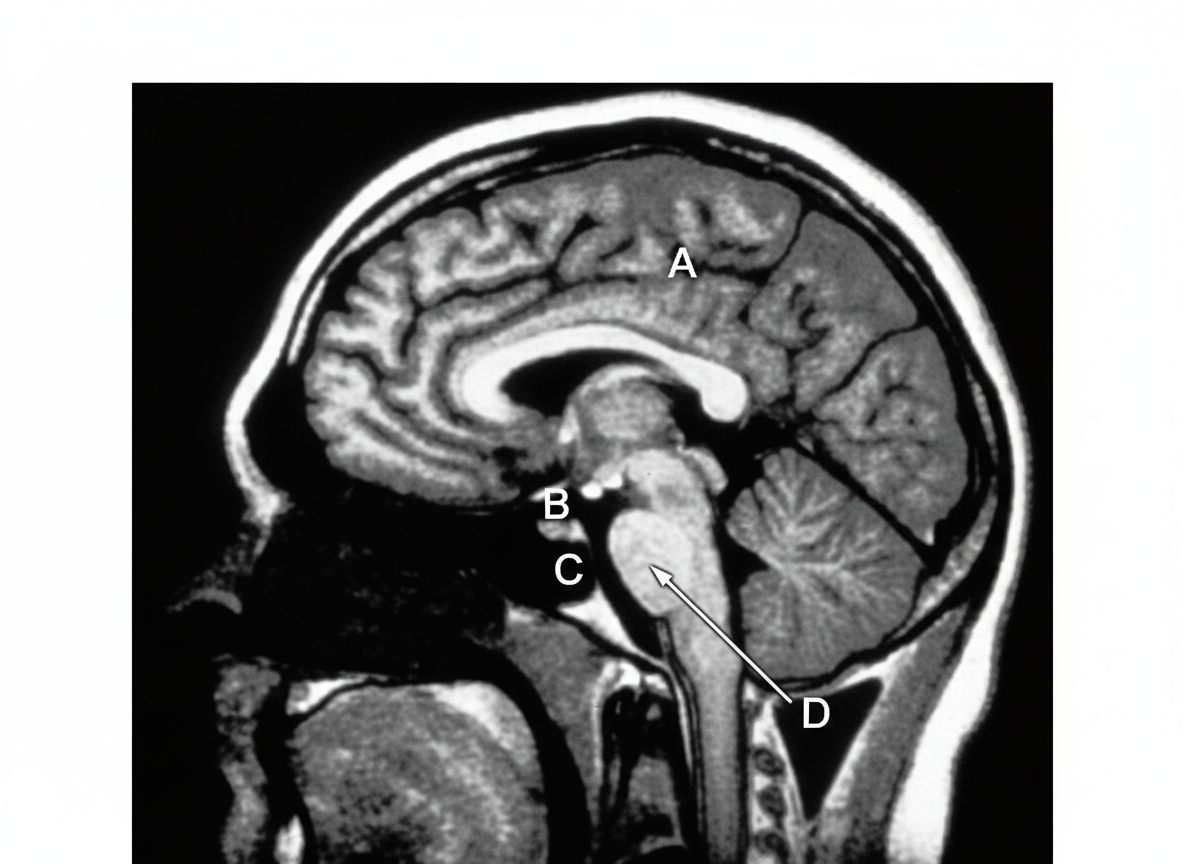
T1 weighted MRI of midsagittal section of the brain is shown. Which among the marked structures contains the two lateral foramina of Luschka?

Which of the following is an oncogenic RNA virus?
Trismus is due to spasm of which muscle?
Which of the following muscles is supplied by two nerves?
All the following structures have a Blood-Brain Barrier except?
What is the name of the junction of the anterior horn and posterior horn of the lateral ventricle?
All of the following structures pass through the superior orbital fissure EXCEPT:
A 'blow-out fracture' of the orbit most commonly involves which part?
Which cranial nerve is the second cranial nerve?
Explanation: ### Explanation **Correct Answer: B. Facial Nerve (CN VII)** The **Facial nerve** provides motor innervation to the **stapedius muscle** via the nerve to stapedius (a branch arising in the facial canal of the temporal bone). The stapedius muscle is responsible for the **stapedial reflex** (acoustic reflex); when exposed to loud sounds, the muscle contracts to pull the stapes away from the oval window, dampening the vibrations of the ossicles [1]. If the facial nerve is injured (commonly seen in Bell’s palsy), the stapedius muscle becomes paralyzed. This loss of the protective dampening mechanism leads to **hyperacusis**, where normal sounds are perceived as uncomfortably loud or distorted. **Analysis of Incorrect Options:** * **A. Hypoglossal (CN XII):** This nerve provides motor supply to the intrinsic and extrinsic muscles of the tongue (except the palatoglossus). Injury results in tongue deviation and atrophy, not auditory symptoms. * **C. Accessory (CN XI):** This nerve innervates the sternocleidomastoid and trapezius muscles. Injury leads to weakness in shrugging shoulders or turning the head. * **D. Vagus (CN X):** The vagus nerve handles parasympathetic outflow, laryngeal/pharyngeal motor control, and visceral sensation. While it has a small sensory branch to the external ear (Arnold’s nerve), it has no role in the middle ear ossicular reflex. **NEET-PG High-Yield Pearls:** * **Tensor Tympani:** This is the other muscle of the middle ear, but it is innervated by the **Mandibular nerve (V3)**. It dampens sounds specifically from chewing. * **Localization:** Hyperacusis in a patient with facial palsy indicates the lesion is **proximal** to the nerve to stapedius (within the facial canal). * **Chorda Tympani:** Often co-injured with the nerve to stapedius, leading to loss of taste on the anterior 2/3 of the tongue.
Explanation: ***D*** - The **fourth ventricle** contains the two lateral **foramina of Luschka** in its lateral recesses, which are **lateral apertures** allowing CSF drainage. - Located in the **posterior fossa** between the brainstem and cerebellum, the fourth ventricle is clearly visible on midsagittal MRI. *A* - This structure does not contain the **foramina of Luschka**, which are specifically located in the lateral recesses of the fourth ventricle. - The **lateral apertures** are anatomically positioned in the posterior fossa, not in this marked region. *B* - This anatomical structure lacks the **lateral recesses** where the foramina of Luschka are located. - The **foramina of Luschka** are exclusive to the fourth ventricle and cannot be found in this marked area. *C* - This region does not house the **fourth ventricle** or its associated **lateral apertures**. - The **foramina of Luschka** are specifically found in the lateral recesses of the fourth ventricle, not in this structure.
Explanation: The core concept tested here is the classification of oncogenic viruses based on their genetic material. While most known oncogenic viruses are DNA viruses, a few specific RNA viruses are strongly associated with human malignancies. **Why Hepatitis C Virus (HCV) is correct:** HCV is a member of the *Flaviviridae* family and is a **single-stranded RNA virus**. It is a major cause of chronic hepatitis, cirrhosis, and **Hepatocellular Carcinoma (HCC)**. Unlike DNA oncogenic viruses, HCV does not integrate its genome into the host cell's DNA. Instead, it promotes oncogenesis through chronic inflammation, oxidative stress, and the action of non-structural proteins (like NS5A) that interfere with cell cycle regulation and apoptosis. **Analysis of Incorrect Options:** * **A. Hepatitis B Virus (HBV):** Although it causes HCC like HCV, HBV is a **partially double-stranded DNA virus** (*Hepadnaviridae*). * **B. Human Papilloma Virus (HPV):** These are **double-stranded DNA viruses**. High-risk strains (16, 18) cause cervical and oropharyngeal cancers by producing E6 and E7 proteins which inhibit p53 and Rb tumor suppressor proteins, respectively. * **C. Epstein-Barr Virus (EBV):** This is a **double-stranded DNA virus** (*Herpesviridae*). It is associated with Burkitt lymphoma, Nasopharyngeal carcinoma, and Hodgkin lymphoma. **High-Yield Clinical Pearls for NEET-PG:** * **Oncogenic RNA Viruses to remember:** Hepatitis C Virus (HCV) and Human T-cell Lymphotropic Virus-1 (HTLV-1) [1]. * **HTLV-1** is the only RNA virus that is directly oncogenic (integrates into the host genome via reverse transcription), causing Adult T-cell Leukemia/Lymphoma [1]. * **HCV** is the only RNA virus among the "Hepatitis" viruses that is commonly associated with chronic carriage leading to malignancy (Hepatitis B is DNA).
Explanation: **Trismus**, commonly known as "lockjaw," refers to the inability to open the mouth due to tonic contraction (spasms) of the muscles of mastication. **Why Medial Pterygoid is the Correct Answer:** The muscles of mastication include the masseter, temporalis, medial pterygoid, and lateral pterygoid. Among these, the **medial pterygoid**, masseter, and temporalis are responsible for **elevating the mandible** (closing the jaw). Spasm of these elevator muscles prevents the jaw from opening. In clinical practice and competitive exams, the medial pterygoid is frequently cited as the primary muscle involved in trismus, especially when associated with dental infections or inferior alveolar nerve blocks. **Analysis of Incorrect Options:** * **A. Orbicularis oris:** This is a muscle of facial expression (supplied by the facial nerve) responsible for closing and puckering the lips, not for jaw movement. * **B. Lateral pterygoid:** This is the only muscle of mastication that **depresses** the mandible (opens the mouth). Spasm of this muscle would theoretically keep the mouth open, not locked shut. * **C. Mentalis:** This is a muscle of facial expression that elevates and protrudes the lower lip (pouting); it has no role in jaw closure. **Clinical Pearls for NEET-PG:** * **Nerve Supply:** All muscles of mastication are supplied by the **mandibular division of the Trigeminal nerve (V3)**. * **Common Causes:** Trismus is a hallmark sign of **Tetanus** (due to *Clostridium tetani* neurotoxin). Other causes include peritonsillar abscess (Quinsy), temporomandibular joint (TMJ) disorders, and impacted third molar infections. * **Key Distinction:** While the masseter is the strongest elevator, the medial pterygoid's involvement is a classic board-style answer for the anatomical basis of trismus.
Explanation: **Explanation:** The **Digastric muscle** is a classic example of a "hybrid" or "composite" muscle because its two bellies are derived from different embryological branchial arches, each carrying its own nerve supply. * **Anterior Belly:** Derived from the **1st branchial arch**, it is supplied by the **nerve to mylohyoid** (a branch of the mandibular nerve, $V_3$). * **Posterior Belly:** Derived from the **2nd branchial arch**, it is supplied by the **digastric branch of the facial nerve** (CN VII). **Analysis of Incorrect Options:** * **Quadriceps:** Supplied solely by the **femoral nerve** ($L_2-L_4$). * **Triceps:** All three heads are supplied by the **radial nerve** ($C_6-C_8$). Note: Occasionally, the medial head receives a branch from the ulnar nerve, but this is an anatomical variation, not the standard supply. * **Flexor Digitorum Superficialis (FDS):** Entirely supplied by the **median nerve**. It is the Flexor Digitorum *Profundus* (FDP) that has a dual supply (median and ulnar nerves). **High-Yield Clinical Pearls for NEET-PG:** * **Other Hybrid Muscles:** Memorize these for exams: * **Pectineus:** Femoral and Obturator nerves. * **Adductor Magnus:** Obturator and Sciatic (tibial part) nerves. * **Flexor Digitorum Profundus:** Median (AIN) and Ulnar nerves. * **Brachialis:** Musculocutaneous and Radial nerves. * **Embryology Link:** Whenever a muscle has a dual nerve supply, it usually signifies a dual embryological origin. For the digastric, the intermediate tendon connects the two developmentally distinct bellies.
Explanation: ### Explanation The **Blood-Brain Barrier (BBB)** is a highly selective semipermeable border of endothelial cells that prevents solutes in the circulating blood from non-selectively crossing into the extracellular fluid of the central nervous system [1]. However, certain specialized areas of the brain, known as **Circumventricular Organs (CVOs)**, lack a BBB to allow for direct sensing of chemical changes in the blood or the release of hormones into the circulation [1], [2]. **Why Option D is Correct:** The **Mammillary bodies** are part of the limbic system (specifically the hypothalamus) and are involved in recollective memory. Unlike CVOs, they possess a **functional blood-brain barrier**. They are not involved in neuroendocrine secretion or systemic chemosensing that would require a fenestrated endothelium. **Why Other Options are Incorrect:** * **Pineal body (Option A):** A sensory CVO that lacks a BBB to secrete melatonin directly into the bloodstream to regulate circadian rhythms. * **Hypophysis cerebri (Option B):** The posterior pituitary (neurohypophysis) lacks a BBB to allow the release of oxytocin and ADH into the systemic circulation [1]. * **Area postrema (Option C):** Located in the floor of the 4th ventricle, it lacks a BBB to act as a "chemoreceptor trigger zone" (CTZ) to detect toxins in the blood and induce vomiting [1]. **High-Yield NEET-PG Pearls:** 1. **Sensory CVOs:** Area postrema, Organum vasculosum of the lamina terminalis (OVLT), and Subfornical organ (SFO) [1]. 2. **Secretory CVOs:** Pineal gland, Posterior pituitary, and Median eminence [1]. 3. **Clinical Correlation:** The lack of BBB in the **Area Postrema** is why chemotherapy drugs often cause severe nausea and vomiting. 4. **Histology:** The BBB is formed by tight junctions between non-fenestrated endothelial cells, the basement membrane, and **astrocyte foot processes**.
Explanation: The **Trigone of the lateral ventricle** (also known as the **Atrium**) is the triangular area where the body, the posterior horn, and the inferior (temporal) horn of the lateral ventricle meet. It is a high-yield anatomical landmark because it contains the largest collection of the choroid plexus, known as the **Glomus choroideum**, which often calcifies with age and is visible on CT scans. ### Why the other options are incorrect: * **Body of lateral ventricle:** This is the central part of the ventricle extending from the interventricular foramen to the trigone. It lies superior to the thalamus and does not represent the junction of the horns. * **Foramen of Monro (Interventricular foramen):** This is the channel that connects the lateral ventricles to the third ventricle. It is located at the anterior end of the body, not at the junction of the horns. * **Cerebral Aqueduct (of Sylvius):** This narrow channel connects the third ventricle to the fourth ventricle within the midbrain. It is not part of the lateral ventricle anatomy. ### NEET-PG Clinical Pearls: * **Glomus Choroideum:** The enlargement of the choroid plexus at the trigone. It is the most common site for intraventricular meningiomas. * **Boundaries:** The trigone is bounded medially by the **Bulb of the posterior horn** (formed by fibers of the Forceps Major) and the **Calcar avis** (produced by the calcarine fissure). * **Hydrocephalus:** Obstruction at the Foramen of Monro leads to unilateral or bilateral dilation of the lateral ventricles, whereas obstruction at the Aqueduct leads to dilation of both lateral and third ventricles.
Explanation: The **Superior Orbital Fissure (SOF)** is a critical communication between the middle cranial fossa and the orbit. It is divided into three parts by the common tendinous ring (Annulus of Zinn). ### **Why Maxillary Nerve is the Correct Answer** The **Maxillary nerve (V2)** does not pass through the SOF. Instead, it exits the middle cranial fossa through the **foramen rotundum** to enter the pterygopalatine fossa. Understanding the exits of the Trigeminal nerve branches is a high-yield NEET-PG concept: * **V1 (Ophthalmic):** Superior Orbital Fissure * **V2 (Maxillary):** Foramen Rotundum * **V3 (Mandibular):** Foramen Ovale ### **Analysis of Incorrect Options** * **Lacrimal nerve:** This is a branch of the Ophthalmic nerve (V1). It passes through the **lateral part** of the SOF (outside the tendinous ring). * **Nasociliary nerve:** This is also a branch of V1. It passes through the **middle part** of the SOF (inside the tendinous ring). * **Inferior ophthalmic vein:** This vein typically passes through the **lower part** of the SOF, though it may occasionally pass through the inferior orbital fissure. ### **High-Yield NEET-PG Clinical Pearls** * **Structures passing OUTSIDE the Annulus of Zinn (Lateral compartment):** **L**acrimal nerve, **F**rontal nerve, **T**rochlear nerve (IV), and **S**uperior ophthalmic vein (Mnemonic: **LFTS**). * **Structures passing INSIDE the Annulus of Zinn (Oculomotor compartment):** Superior and inferior divisions of **Oculomotor nerve (III)**, **Abducens nerve (VI)**, and **Nasociliary nerve**. * **Superior Orbital Fissure Syndrome:** Characterized by ophthalmoplegia (palsy of CN III, IV, VI) and anesthesia in the V1 distribution, but with normal vision (as the Optic nerve is spared).
Explanation: Explanation: A **blow-out fracture** occurs when a blunt object (larger than the orbital rim, such as a tennis ball or fist) strikes the orbit. The impact increases intraorbital pressure, which is transmitted to the orbital walls. The fracture occurs at the weakest points to "decompress" the orbit. **1. Why the Floor is Correct:** The **orbital floor** is the most common site of a blow-out fracture because it is composed of the thin orbital plate of the **maxilla**. Specifically, the area medial to the infraorbital groove is the thinnest part of the entire orbit. When the floor fractures, orbital contents (fat and the **inferior rectus muscle**) can herniate into the **maxillary sinus**, leading to characteristic clinical signs like enophthalmos and diplopia on upward gaze. **2. Analysis of Incorrect Options:** * **Apex:** This is the strongest part of the orbit where the extraocular muscles originate. It requires massive trauma and is rarely involved in isolated blow-out injuries. * **Lateral Wall:** This is the thickest and strongest wall of the orbit, formed by the zygomatic bone and the greater wing of the sphenoid. * **Base:** This refers to the orbital rim, which is thick and reinforced. By definition, a "true" blow-out fracture involves the internal walls while the orbital rim (base) remains intact. **High-Yield Clinical Pearls for NEET-PG:** * **Second most common site:** The **medial wall** (lamina papyracea of the ethmoid bone). * **Clinical Sign:** Diplopia on upward gaze due to entrapment of the **Inferior Rectus muscle**. * **Nerve Involvement:** Anesthesia of the cheek and upper gum due to injury to the **Infraorbital nerve**. * **Radiology:** The **"Teardrop sign"** on a Waters’ view X-ray (herniated orbital fat in the maxillary sinus).
Explanation: **Explanation:** The cranial nerves are a set of 12 paired nerves that emerge directly from the brain and brainstem, numbered according to their rostrocaudal (front-to-back) exit from the brain. The **Optic nerve (CN II)** is the correct answer as it is the second cranial nerve, responsible for transmitting visual information from the retina to the brain. **Analysis of Options:** * **Optic nerve (CN II):** It originates from the ganglion cells of the retina and enters the middle cranial fossa via the optic canal [1]. It is unique because it is technically an extension of the forebrain (diencephalon) rather than a peripheral nerve. * **Abducens nerve (CN VI):** This is the sixth cranial nerve. It emerges from the pontomedullary junction and supplies the lateral rectus muscle for eye abduction. * **Trigeminal nerve (CN V):** This is the fifth and largest cranial nerve. it emerges from the pons and provides sensory innervation to the face and motor innervation to the muscles of mastication. * **Oculomotor nerve (CN III):** This is the third cranial nerve. It emerges from the midbrain and controls most of the extraocular muscles and pupillary constriction. **High-Yield Clinical Pearls for NEET-PG:** * **Myelination:** Unlike most peripheral nerves myelinated by Schwann cells, the Optic nerve is myelinated by **oligodendrocytes**, making it susceptible to Multiple Sclerosis. * **Meningeal Covering:** Since it is an outgrowth of the brain, it is covered by all three layers of meninges (dura, arachnoid, and pia mater). * **Clinical Sign:** A lesion in the optic nerve leads to an **Ipsilateral Anopsia** and a loss of the direct light reflex in the affected eye [1].
Organization of the Nervous System
Practice Questions
Spinal Cord Anatomy
Practice Questions
Brainstem Anatomy
Practice Questions
Cerebellum
Practice Questions
Diencephalon
Practice Questions
Cerebral Cortex
Practice Questions
Basal Ganglia
Practice Questions
Limbic System
Practice Questions
Cranial Nerves
Practice Questions
Autonomic Nervous System
Practice Questions
Neural Pathways and Tracts
Practice Questions
Neurovascular Anatomy
Practice Questions
Get full access to all questions, explanations, and performance tracking.
Start For Free